
Page 208 - Color_Atlas_of_Physiology_5th_Ed._-_A._Despopoulos_2003
P. 208
Cardiac Impulse Generation and Conduction II
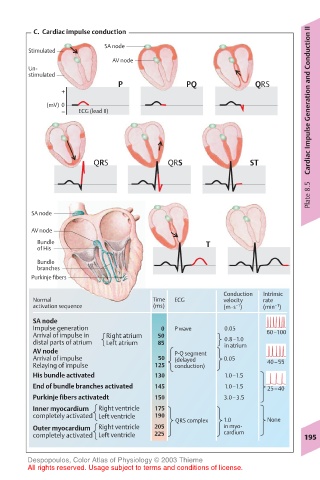
C. Cardiac impulse conduction
SA node
Stimulated
AV node
Un-
stimulated
P PQ QRS
+
(mV) 0
– ECG (lead II)
QRS QRS ST
Plate 8.5
SA node
AV node
Bundle T
of His
Bundle
branches
Purkinje fibers
Conduction Intrinsic
Normal Time ECG velocity rate
activation sequence (ms) (m·s ) (min )
–1
–1
SA node
Impulse generation 0 P wave 0.05 60–100
Arrival of impulse in Right atrium 50 0.8–1.0
distal parts of atrium Left atrium 85 in atrium
AV node P-Q segment
Arrival of impulse 50 (delayed 0.05 40–55
Relaying of impulse 125 conduction)
His bundle activated 130 1.0–1.5
End of bundle branches activated 145 1.0–1.5 25–40
Purkinje fibers activatedt 150 3.0–3.5
Inner myocardium Right ventricle 175
completely activated Left ventricle 190
QRS complex 1.0 None
Outer myocardium Right ventricle 205 in myo-
completely activated Left ventricle 225 cardium 195
Despopoulos, Color Atlas of Physiology © 2003 Thieme
All rights reserved. Usage subject to terms and conditions of license.