
Page 206 - Color_Atlas_of_Physiology_5th_Ed._-_A._Despopoulos_2003
P. 206
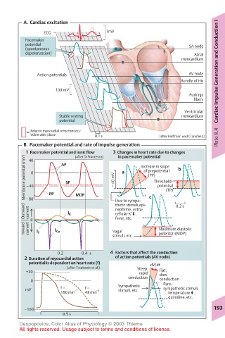
A.  Cardiac excitation
               ECG                 1mV
         Pacemaker
         potential                                            SA node
         (spontaneous
         depolarization)                                       Atrial
                                                           myocardium
             Action potentials                               AV node   Cardiac Impulse Generation and Conduction I
                                                           Bundle of His
                   100 mV
                                                              Purkinje
                                                               fibers

                                                            Ventricular
                    Stable resting                         myocardium
                    potential
           Relative myocardial refractoriness:
           Vulnerable phase    0.1 s                (after Hoffman und Cranefield)  Plate 8.4
         B.  Pacemaker potential and rate of impulse generation
         1  Pacemaker potential and ionic flow  3  Changes in heart rate due to changes
                                           in pacemaker potential
                         (after Di Francesco)
         Membrane potential (mV)  –40 0  PP  SP  – 40 mV  a  Increase in slope  b
          40
                     AP
                                               of prepotential
                                                       (PP)
                                                  Threshold-
                                                   potential
                                                      (TP)
                                      thetic stimuli,epi-
         Outward  –80  I K  MDP       Due to sympa-  c   0.2 s
          current
                                      nephrine, extra-
                                           +
                                      cellular K
                                            ,
           0                          fever, etc.
         Inward  current  I Ca       Vagal          Maximum diastolic
             I f
                                     stimuli, etc.  potential (MDP)
                   0.2     0.4 s    4  Factors that affect the conduction
         2 Duration of myocardial action        of action potentials (AV node)
              potential is dependent on heart rate (f)  dV/dt
                       (after Trautwein et al.)  Steep:
         +30                                  rapid  Flat:
                                          conduction  slow
           0                                        conduction
                                       Sympathetic   Para-
         mV         f =  –1  f =  –1   stimuli, etc.  sympathetic stimuli,
                    160min  48min                          temperature     ,
                                                           quinidine, etc.
         –100                                                        193
                     0.5 s
       Despopoulos, Color Atlas of Physiology © 2003 Thieme
       All rights reserved. Usage subject to terms and conditions of license.