
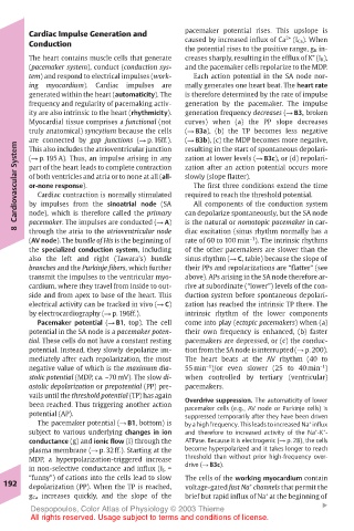
Page 205 - Color_Atlas_of_Physiology_5th_Ed._-_A._Despopoulos_2003
P. 205
Cardiac Impulse Generation and  pacemaker potential rises. This upslope is
                                                           2+
       Conduction                      caused by increased influx of Ca  (I Ca). When
                                       the potential rises to the positive range, g K in-
                                                                 +
       The heart contains muscle cells that generate  creases sharply, resulting in the efflux of K (I K),
       (pacemaker system), conduct (conduction sys-  and the pacemaker cells repolarize to the MDP.
       tem) and respond to electrical impulses (work-  Each action potential in the SA node nor-
       ing  myocardium).  Cardiac  impulses  are  mally generates one heart beat. The heart rate
       generated within the heart (automaticity). The  is therefore determined by the rate of impulse
       frequency and regularity of pacemaking activ-  generation by the pacemaker. The impulse
       ity are also intrinsic to the heart (rhythmicity).  generation frequency decreases (! B3, broken
       Myocardial tissue comprises a functional (not  curves) when (a) the PP slope decreases
       truly anatomical) syncytium because the cells  (! B3a), (b) the TP becomes less negative
       are connected by gap junctions (! p. 16ff.).  (! B3b), (c) the MDP becomes more negative,
    Cardiovascular System  part of the heart leads to complete contraction  zation after an action potential occurs more
                                       resulting in the start of spontaneous depolari-
       This also includes the atrioventricular junction
                                       zation at lower levels (! B3c), or (d) repolari-
       (! p. 195 A). Thus, an impulse arising in any
       of both ventricles and atria or to none at all (all-
                                       slowly (slope flatter).
                                        The first three conditions extend the time
       or-none response).
                                       required to reach the threshold potential.
         Cardiac contraction is normally stimulated
                                        All components of the conduction system
       by impulses from the sinoatrial node (SA
                                       is the natural or nomotopic pacemaker in car-
       pacemaker. The impulses are conducted (! A)
    8  node), which is therefore called the primary  can depolarize spontaneously, but the SA node
                                       diac excitation (sinus rhythm normally has a
       through the atria to the atrioventricular node
                                                    –1
       (AV node). The bundle of His is the beginning of  rate of 60 to 100 min ). The intrinsic rhythms
       the specialized conduction system, including  of the other pacemakers are slower than the
       also the left and right (Tawara’s) bundle  sinus rhythm (! C, table) because the slope of
       branches and the Purkinje fibers, which further  their PPs and repolarizations are “flatter” (see
       transmit the impulses to the ventricular myo-  above). APs arising in the SA node therefore ar-
       cardium, where they travel from inside to out-  rive at subordinate (“lower”) levels of the con-
       side and from apex to base of the heart. This  duction system before spontaneous depolari-
       electrical activity can be tracked in vivo (! C)  zation has reached the intrinsic TP there. The
       by electrocardiography (! p. 196ff.).  intrinsic rhythm of the lower components
         Pacemaker potential (! B1, top). The cell  come into play (ectopic pacemakers) when (a)
       potential in the SA node is a pacemaker poten-  their own frequency is enhanced, (b) faster
       tial. These cells do not have a constant resting  pacemakers are depressed, or (c) the conduc-
       potential. Instead, they slowly depolarize im-  tion from the SA node is interrupted (! p. 200).
       mediately after each repolarization, the most  The heart beats at the AV rhythm (40 to
                                           –1
                                                                  –1
       negative value of which is the maximum dia-  55 min )/or even slower (25 to 40 min )
       stolic potential (MDP, ca. –70 mV). The slow di-  when controlled by tertiary (ventricular)
       astolic depolarization or prepotential (PP) pre-  pacemakers.
       vails until the threshold potential (TP) has again  Overdrive suppression. The automaticity of lower
       been reached. Thus triggering another action  pacemaker cells (e.g., AV node or Purkinje cells) is
       potential (AP).                 suppressed temporarily after they have been driven
         The pacemaker potential (! B1, bottom) is  by a high frequency. This leads to increased Na influx
                                                                +
                                                                 +
       subject to various underlying changes in ion  and therefore to increased activity of the Na -K - +
       conductance (g) and ionic flow (I) through the  ATPase. Because it is electrogenic (! p. 28), the cells
       plasma membrane (! p. 32ff.). Starting at the  become hyperpolarized and it takes longer to reach
       MDP, a hyperpolarization-triggered increase  threshold than without prior high-frequency over-
                                       drive (! B3c).
       in non-selective conductance and influx (I f, =
       “funny”) of cations into the cells lead to slow  The cells of the working myocardium contain
  192  depolarization (PP). When the TP is reached,  voltage-gated fast Na channels that permit the
                                                    +
       g Ca increases quickly, and the slope of the  brief but rapid influx of Na at the beginning of
                                                       +
                                                                   !
       Despopoulos, Color Atlas of Physiology © 2003 Thieme
       All rights reserved. Usage subject to terms and conditions of license.