
Page 219 - Color_Atlas_of_Physiology_5th_Ed._-_A._Despopoulos_2003
P. 219
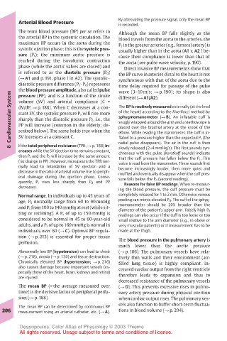
Arterial Blood Pressure         By attenuating the pressure signal, only the mean BP
                                       is recorded.
       The term blood pressure (BP) per se refers to  Although the mean BP falls slightly as the
       the arterial BP in the systemic circulation. The  blood travels from the aorta to the arteries, the
       maximum BP occurs in the aorta during the  P s in the greater arteries (e.g., femoral artery) is
       systolic ejection phase; this is the systolic pres-  usually higher than in the aorta (A1 v. A2 ) be-
       sure (P s); the minimum aortic pressure is  cause their compliance is lower than that of
       reached during the isovolumic contraction  the aorta (see pulse wave velocity, p. 190).
       phase (while the aortic valves are closed) and  Direct invasive BP measurements show that
       is referred to as the diastolic pressure (P d)  the BP curve in arteries distal to the heart is not
       (! A1 and p. 191, phase I in A2). The systolic–  synchronous with that of the aorta due to the
       diastolic pressure difference (P s–P d) represents  time delay required for passage of the pulse
       the blood pressure amplitude, also called pulse  wave (3–10 m/s; ! p. 190); its shape is also
    Cardiovascular System  dV/dP, ! p. 188). When C decreases at a con-  The BP is routinely measured externally (at the level
       pressure (PP), and is a function of the stroke
                                       different (! A1/A2).
       volume (SV) and arterial compliance (C =
                                       of the heart) according to the Riva-Rocci method by
       stant SV, the systolic pressure P s will rise more
                                       sphygmomanometer (! B). An inflatable cuff is
       sharply than the diastolic pressure P d, i.e., the
                                       snugly wrapped around the arm and a stethoscope is
       PP will increase (common in the elderly; de-
                                       placed over the brachial artery at the crook of the
       scribed below). The same holds true when the
                                       flated to a pressure higher than the expected P s (the
                                       radial pulse disappears). The air in the cuff is then
       If the total peripheral resistance (TPR, ! p. 188) in-
    8  SV increases at a constant C.   elbow. While reading the manometer, the cuff is in-
                                       slowly released (2–4 mmHg/s). The first sounds syn-
       creases while the SV ejection time remains constant,  chronous with the pulse (Korotkoff sounds) indicate
       then P s and the P d will increase by the same amount  that the cuff pressure has fallen below the P s. This
       (no change in PP). However, increases in the TPR nor-  value is read from the manometer. These sounds first
       mally lead to retardation of SV ejection and a  become increasingly louder, then more quiet and
       decrease in the ratio of arterial volume rise to periph-  muffled and eventually disappear when the cuff pres-
       eral drainage during the ejection phase. Conse-  sure falls below the P d (second reading).
       quently, P s rises less sharply than P d and PP  Reasons for false BP readings. When re-measur-
       decreases.                      ing the blood pressure, the cuff pressure must be
       Normal range. In individuals up to 45 years of  completely released for 1 to 2 min. Otherwise venous
       age, P d normally range from 60 to 90 mmHg  pooling can mimic elevated P d. The cuff of the sphyg-
       and P s from 100 to 140 mmHg at rest (while sit-  momanometer should be 20% broader than the
       ting or reclining). A P s of up to 150 mmHg is  diameter of the patient’s upper arm. Falsely high P d
                                       readings can also occur if the cuff is too loose or too
       considered to be normal in 45 to 60-year-old  small relative to the arm diameter (e.g., in obese or
       adults, and a P s of up to 160 mmHg is normal in  very muscular patients) or if measurement has to be
       individuals over 60 (! C). Optimal BP regula-  made at the thigh.
       tion (! p. 212) is essential for proper tissue
       perfusion.                      The blood pressure in the pulmonary artery is
                                       much  lower  than  the  aortic  pressure
       Abnormally low BP (hypotension) can lead to shock  (! p. 186). The pulmonary vessels have rela-
       (! p. 218), anoxia (! p. 130) and tissue destruction.  tively thin walls and their environment (air-
       Chronically elevated BP (hypertension; ! p. 216)  filled lung tissue) is highly compliant. In-
       also causes damage because important vessels (es-  creased cardiac output from the right ventricle
       pecially those of the heart, brain, kidneys and retina)
       are injured.                    therefore leads to expansion and thus to
                                       decreased resistance of the pulmonary vessels
       The mean BP (= the average measured over  (! D). This prevents excessive rises in pulmo-
       time) is the decisive factor of peripheral perfu-  nary artery pressure during physical exertion
       sion (! p. 188).                when cardiac output rises. The pulmonary ves-
                                       sels also function to buffer short-term fluctua-
       The mean BP can be determined by continuous BP
  206  measurement using an arterial catheter, etc. (! A).  tions in blood volume (! p. 204).
       Despopoulos, Color Atlas of Physiology © 2003 Thieme
       All rights reserved. Usage subject to terms and conditions of license.