
Page 372 - Color_Atlas_of_Physiology_5th_Ed._-_A._Despopoulos_2003
P. 372
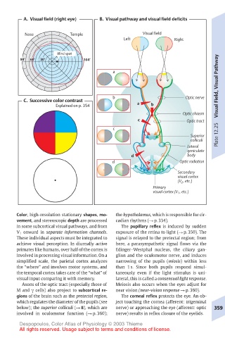
A.  Visual field (right eye)  B.  Visual pathway and visual field deficits

         Nose          Temple                  Visual field
                                         Left            Right

                   Blind spot
                                                                       Visual Field, Visual Pathway
       90°  60°  30°        104°
                                     a


                                     b                      Optic nerve
         C.  Successive color contrast
                   Explained on p. 354       a   b
                                                            Optic chiasm
                                             c               Optic tract
                                     c
                                                              Superior  Plate 12.25
                                                              colliculi
                                                             Lateral
                                                             geniculate
                                           d                 body
                                     d                     Optic radiation
                                                          Secondary
                                                          visual cortex
                                                          (V 2 , etc.)
                                                  Primary
                                                  visual cortex (V 1 , etc.)


      Color, high-resolution stationary shapes, mo-  the hypothalamus, which is responsible for cir-
      vement, and stereoscopic depth are processed  cadian rhythms (! p. 334).
      in some subcortical visual pathways, and from  The pupillary reflex is induced by sudden
      V 1 onward in separate information channels.  exposure of the retina to light (! p. 350). The
      These individual aspects must be integrated to  signal is relayed to the pretectal region; from
      achieve visual perception. In diurnally active  here, a parasympathetic signal flows via the
      primates like humans, over half of the cortex is  Edinger–Westphal nucleus, the ciliary gan-
      involved in processing visual information. On a  glion and the oculomotor nerve, and induces
      simplified scale, the parietal cortex analyzes  narrowing of the pupils (miosis) within less
      the “where” and involves motor systems, and  than 1 s. Since both pupils respond simul-
      the temporal cortex takes care of the “what” of  taneously even if the light stimulus is uni-
      visual input comparing it with memory.  lateral, this is called a consensual light response.
        Axons of the optic tract (especially those of  Meiosis also occurs when the eyes adjust for
      M and γ cells) also project to subcortical re-  near vision (near-vision response ! p. 360).
      gions of the brain such as the pretectal region,  The corneal reflex protects the eye. An ob-
      which regulates the diameter of the pupils (see  ject touching the cornea (afferent: trigeminal
      below); the superior colliculi (! B), which are  nerve) or approaching the eye (afferent: optic  359
      involved in oculomotor function (! p. 360);  nerve) results in reflex closure of the eyelids.
       Despopoulos, Color Atlas of Physiology © 2003 Thieme
       All rights reserved. Usage subject to terms and conditions of license.