
Page 1148 - Hall et al (2015) Principles of Critical Care-McGraw-Hill
P. 1148
CHAPTER 86: Intracranial Pressure: Monitoring and Management 787
fixed space of the calvarium must be carefully regulated by many mecha- The structures between the brain surface and the inner skull,
nisms in order to be maintained within a physiologic range. Disruption the meningeal layers, are important in identifying and maintaining
of these mechanisms through trauma, space-occupying lesions, or normal ICP. The most important of these is the subarachnoid space
edema leads to dysregulation of the delicate balance required to main- where the arachnoid villi conduct cerebrospinal fluid (CSF) from the
tain normal pressure that results in significant neurologic and systemic subarachnoid space to the venous sinuses. If these granulations are
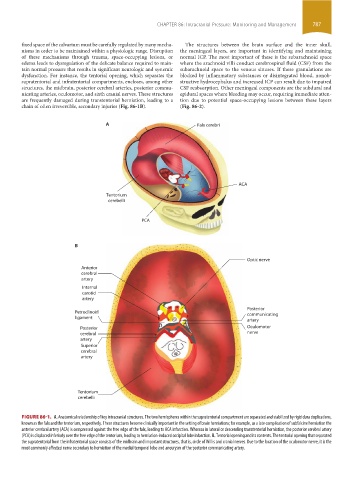
dysfunction. For instance, the tentorial opening, which separates the blocked by inflammatory substances or disintegrated blood, nonob-
supratentorial and infratentorial compartments, encloses, among other structive hydrocephalus and increased ICP can result due to impaired
structures, the midbrain, posterior cerebral arteries, posterior commu- CSF reabsorption. Other meningeal components are the subdural and
nicating arteries, oculomotor, and sixth cranial nerves. These structures epidural spaces where bleeding may occur, requiring immediate atten-
are frequently damaged during transtentorial herniation, leading to a tion due to potential space-occupying lesions between these layers
chain of often irreversible, secondary injuries (Fig. 86-1B). (Fig. 86-2).
A Falx cerebri
ACA
Tentorium
cerebelli
PCA
B
Optic nerve
Anterior
cerebral
artery
Internal
carotid
artery
Posterior
Petroclinoid communicating
ligament
artery
Posterior Oculomotor
cerebral nerve
artery
Superior
cerebral
artery
Tentorium
cerebelli
FIGURE 86-1. A. Anatomical relationship of key intracranial structures. The two hemispheres within the supratentorial compartment are separated and stabilized by rigid dura duplications,
known as the falx and the tentorium, respectively. These structures become clinically important in the setting of brain herniations; for example, as a late complication of subfalcine herniation the
anterior cerebral artery (ACA) is compressed against the free edge of the falx, leading to ACA infarction. Whereas in lateral or descending transtentorial herniation, the posterior cerebral artery
(PCA) is displaced inferiorly over the free edge of the tentorium, leading to herniation-induced occipital lobe infarction. B. Tentorial opening and its contents. The tentorial opening that separated
the supratentorial from the infratentorial space consists of the midbrain and important structures, that is, circle of Willis and cranial nerves. Due to the location of the oculomotor nerve, it is the
most commonly affected nerve secondary to herniation of the medial temporal lobe and aneurysm of the posterior communicating artery.
section06.indd 787 1/23/2015 12:55:43 PM