
Page 1150 - Hall et al (2015) Principles of Critical Care-McGraw-Hill
P. 1150
CHAPTER 86: Intracranial Pressure: Monitoring and Management 789
TABLE 86-1 Causes of Elevated Intracranial Pressure Simultaneous ECG
Primary (Intracranial) Secondary (Extracranial)
200 40
Nontraumatic Airway obstruction Arterial pressure
Intracranial hemorrhages (parenchymal, Hypoventilation
subarachnoid, subdural, epidural) Hypoxia 150 30
Ischemic infarction Hypercarbia
Hydrocephalus (communicating and Head position or posture Systemic arterial pressure Intracranial pressure
noncommunicating) Venous outflow obstruction
Brain edema Hyperpyrexia 100 20
Brain tumor Hyponatremia
Status epilepticus Agitation, pain
Cerebral venous thrombosis Diabetic ketoacidosis 50 10
Cerebral vasospasm Eclampsia or hypertensive encephalopathy Intracranial pressure
Infection (ie, encephalitis, meningitis, Convulsive or nonconvulsive seizure
abscess, etc) Increased intrathoracic or intra-abdominal 0 0
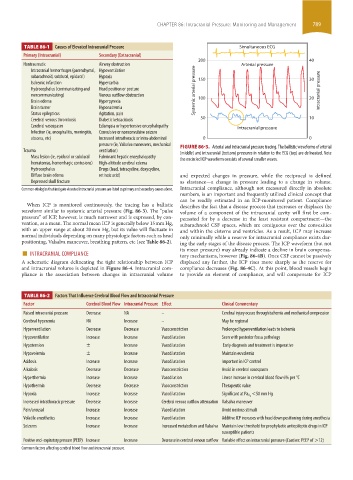
pressure (ie, Valsalva maneuvers, mechanical FIGURE 86-3. Arterial and intracranial pressure tracing. The ballistic waveforms of arterial
Trauma ventilation) (middle) and intracranial (bottom) pressures in relation to the ECG (top) are delineated. Note
Mass lesion (ie, epidural or subdural Fulminant hepatic encephalopathy the encircled ICP waveform consists of several smaller waves.
hematomas, hemorrhagic contusions) High-altitude cerebral edema
Hydrocephalus Drugs (lead, tetracycline, doxycycline,
Diffuse brain edema retinoic acid) and expected changes in pressure, while the reciprocal is defined
Depressed skull fracture as elastance—a change in pressure leading to a change in volume.
Common etiologies that instigate elevated intracranial pressure are listed as primary and secondary causes above. Intracranial compliance, although not measured directly in absolute
numbers, is an important and frequently utilized clinical concept that
can be readily estimated in an ICP-monitored patient. Compliance
When ICP is monitored continuously, the tracing has a ballistic describes the fact that a disease process that increases or displaces the
waveform similar to systemic arterial pressure (Fig. 86-3). The “pulse volume of a component of the intracranial cavity will first be com-
pressure” of ICP, however, is much narrower and is expressed, by con- pensated for by a decrease in the least resistant compartment—the
vention, as a mean. The normal mean ICP is generally below 15 mm Hg, subarachnoid CSF spaces, which are contiguous over the convexities
with an upper range at about 20 mm Hg, but its value will fluctuate in and within the cisterns and ventricles. As a result, ICP may increase
normal individuals depending on many physiologic factors such as head only minimally while a reserve for intracranial compliance exists dur-
positioning, Valsalva maneuver, breathing pattern, etc (see Table 86-2). ing the early stages of the disease process. The ICP waveform (but not
■ INTRACRANIAL COMPLIANCE its mean pressure) may already indicate a decline in brain compensa-
tory mechanisms, however (Fig. 86-4B). Once CSF cannot be passively
A schematic diagram delineating the tight relationship between ICP displaced any further, the ICP rises more sharply as the reserve for
and intracranial volume is depicted in Figure 86-4. Intracranial com- compliance decreases (Fig. 86-4C). At this point, blood vessels begin
pliance is the association between changes in intracranial volume to provide an element of compliance, and will compensate for ICP
TABLE 86-2 Factors That Influence Cerebral Blood Flow and Intracranial Pressure
Factor Cerebral Blood Flow Intracranial Pressure Effect Clinical Commentary
Raised intracranial pressure Decrease NA – Cerebral injury occurs through ischemia and mechanical compression
Cerebral hyperemia NA Increase – May be regional
Hyperventilation Decrease Decrease Vasoconstriction Prolonged hyperventilation leads to ischemia
Hypoventilation Increase Increase Vasodilatation Seen with posterior fossa pathology
Hypotension ± Increase Vasodilatation Early diagnosis and treatment is imperative
Hypovolemia ± Increase Vasodilatation Maintain euvolemia
Acidosis Increase Increase Vasodilatation Important in ICP control
Alkalosis Decrease Decrease Vasoconstriction Avoid in cerebral vasospasm
Hyperthermia Increase Increase Vasodilation Linear increase in cerebral blood flow 6% per °C
Hypothermia Decrease Decrease Vasoconstriction Therapeutic value
Hypoxia Increase Increase Vasodilatation Significant at Pa O 2 <50 mm Hg
Increased intrathoracic pressure Decrease Increase Cerebral venous outflow attenuation Valsalva maneuver
Pain/arousal Increase Increase Vasodilatation Avoid noxious stimuli
Volatile anesthetics Increase Increase Vasodilatation Additive ICP increases with head down positioning during anesthesia
Seizures Increase Increase Increased metabolism and Valsalva Maintain low threshold for prophylactic antiepileptic drugs in ICP
susceptible patients
Positive end- expiratory pressure (PEEP) Increase Increase Decrease in cerebral venous outflow Variable effect on intracranial pressure (Caution: PEEP of >12)
Common factors affecting cerebral blood flow and intracranial pressure.
section06.indd 789 1/23/2015 12:55:46 PM