
Page 1153 - Hall et al (2015) Principles of Critical Care-McGraw-Hill
P. 1153
792 PART 6: Neurologic Disorders
Autoregulatory curve
Passive Maximum Zone of normal Maximum
Collapse Dilatation Autoregulation Constriction
Cerebral blood flow (mL/100 g/min) 75 4.0 Cerebral blood volume (mL/100 g)
100
50
25
0
25 50 75 100 125 150 175 200
Cerebral perfusion pressure (mm Hg)
Legend:
Normal autoregulation
Cerebrovascular resistance
Cerebral blood volume
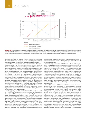
FIGURE 86-7. Autoregulatory curve. With intact cerebral autoregulation the cerebral blood flow is maintained constant over a wide range of cerebral perfusion pressures (50-150 mm Hg;
solid red line). Outside of this pressure range, cerebral arterioles collapse at very low CPP or at very high pressures abnormally (breakthrough) constricts. Abnormal autoregulation, commonly
present in injured brain, is the complete dependence of cerebral perfusion on systemic arterial pressures, an abnormality that has important consequences on intracranial pressure.
decreased blood flow. For example, a CBF of 18 to 23 mL/100 g/min can cerebral arterial tone is also regulated by sympathetic input leading to
be tolerated for 2 weeks, as opposed to 10 to 12 mL/100 g/min for 3 hours mild tonic vasoconstriction and allowing for higher limits to be reached
and 8 mL/100 g/min for only 1 hour before neuronal death ensues. on the regulation curve. 7
In addition to autoregulatory cerebral vascular failure, the injured Clinically important factors that influence CBF and ICP are pre-
brain also suffers from uncoupling of cerebral metabolism. In normal sented in Table 86-2. Control of these factors constitutes the basis for
brain, cerebral metabolic demand and regional blood flow fluctuate in a much of the medical management of raised ICP in brain injury. For
proportional manner. Neural activation leads to increased cerebral met- example, CVR changes linearly within a Pa CO 2 range between 20 and
abolic activity, which increases the demand for glucose and oxygen and 80 mm Hg. As a result, Pa CO 2 and its manipulation have a dramatic
is met by local increases in CBF. This phenomenon is called metabolic effect on CBF and ICP even when CPP is held constant by alterations
autoregulation, with the interaction between metabolic fluctuation and in MAP. As an example, inhalation of low CO concentrations (5%-
2
alterations in ICP intimately intertwined at the precapillary level. The 7%) can double CBF through changes in extracellular pH that lead to
close coupling between metabolic supply and demand can be monitored vasodilation of cerebral vasculature and a resultant increase in ICP. To
and clinically applied to managing brain-injured patients by correlating the contrary, low CO created by hyperventilation results in vasocon-
2
cerebral metabolic rate of oxygen consumption (CMRO ) and the arte- striction and lowered ICP and can ultimately result in brain ischemia
2
riovenous difference in oxygen saturation (AVDO ) as expressed by the due to prolonged vasoconstriction. Changes in Pa O 2 also affect CBF
2
Fick equation: CMRO = CBF × AVDO or AVDO = CMRO /CBF. In when values fall to less than ~50 mm Hg, which is demonstrated
2
2
2
2
healthy brain parenchyma, AVDO is constant, and changes in demand in Figure 86-9.
2
are met by changes in CBF by adjusting local CVR. In the traumatized Although the optimal CPP for each individual may vary, it is recom-
brain, however, mismatch of supply and demand in the face of preexist- mended that in healthy subjects CPP be maintained above 50 mm Hg
ing abnormal pressure autoregulation can lead to AVDO that may be to avoid ischemia and less than 110 mm Hg to avoid breakthrough
2
either higher or lower than necessary. hyperperfusion and cerebral edema. Current traumatic brain injury
Under physiologic conditions, a MAP of 80 to 100 mm Hg and an ICP (TBI) guidelines suggest that the optimal CPP in head trauma resides
of 5 to 10 mm Hg can lead to a CPP of 70 to 85 mm Hg. However, the between 50 and 70 mm Hg as CPP levels above 70 mm Hg failed to
9
true regional CPP may vary by as much as 27 mm Hg from measure- improve outcome and caused a fivefold increase in acute respiratory
ments utilizing global MAP and ICP values. Obtaining accurate MAP distress syndrome. The presence of a U-shaped curve between CPP
6,10
8
measurements for CPP determination requires the placement of an arte- and measures of intact cerebral autoregulation, such as PRx and Mx
rial pressure catheter, with its transducer at the level of the foramen of (see below), identifies that both inadequate and excessive CPP are
Monro, which approximates to the level of external auditory meatus (see associated with failure of autoregulatory mechanisms. When CPP drops
Fig. 86-8). The ICP should be measured in units of mm Hg to accurately below 40, autoregulation fails and blood vessels collapse (Fig. 86-7),
calculate CPP. In the normal brain, CBF is constant as long as the MAP lowering both intracranial blood volume and CBF. Conversely, CPP
is maintained between 50 and 150 mm Hg. The local regulation of arte- sustained above 110 mm Hg overcomes mechanisms of autoregulation
rial vascular resistance is affected by CO , O , pH/lactate levels, adenos- and hyperperfuses the brain due to passive, irreversible dilation of arte-
2
2
ine, nitric oxide, and other components. Neurogenic regulation of the rioles with resultant elevation in brain swelling and ICP. Maintenance
section06.indd 792 1/23/2015 12:55:49 PM