
Page 1149 - Hall et al (2015) Principles of Critical Care-McGraw-Hill
P. 1149
788 PART 6: Neurologic Disorders
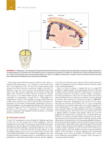
Epidural Arachnoid
Pia mater
Dura Skull
mater
Skin
Subdural
Brain
FIGURE 86-2. Meningeal layers. The cerebrospinal fluid compartments located between the brain surface and inner skull are clinically described as subarachnoid, subdural, and (functionally
nonexisting) epidural spaces. Arachnoid villi within the subarachnoid space have the important role of continuous CSF absorption and, if obstructed due to infection or disintegrated blood prod-
ucts, communicating hydrocephalus and eventually elevated intracranial pressure will occur. The subdural and epidural spaces are important in that blood and fluid may track into and expand
these potential spaces in the setting of trauma or ruptured vascular malformations.
The average volume within the cranium is 1500 mL, with ~88% con- of the ICP pulse waveform is the response of ICP to that increment of
sisting of brain parenchyma, ~7.5% composed of intracranial blood, volume. The properties of ICP wave should therefore be directly related
and ~4.5% composed of CSF. The sum of the partial pressures and to the craniospinal elastance. 2
1
volumes from these three main components is equal to the total ICP. There is no level I evidence to support the use of a single ICP
Therefore, when one volume increases (eg, intraparenchymal brain threshold to initiate therapy. The recommended critical ICP elevation
tumor), the other volumes compensate for the pressure change and in adults at which treatment should be initiated (Level II evidence) is
reduce their combined intracranial volumes to keep ICP constant. 20 mm Hg sustained for more than 5 minutes. Failure of compensa-
3
This is known as the Monro-Kellie doctrine. Frequent mechanisms tory brain mechanisms to reduce ICP to normal values will result in
responsible for maintaining a normal ICP (ie, <20 mm Hg) are a intracranial hypertension and its clinical sequelae. Common etiologies
compensatory increase in CSF absorption, drainage of blood from the for primary and secondary ICP elevations are listed in Table 86-1.
cerebral venous systems, and a shift of CSF from the cranial subarach- Numerous studies have demonstrated that elevated ICP is associ-
noid space into the spinal (intraforaminal) compartment. Because of ated with poor outcome and, therefore, that ICP control and pressure
skull noncompliance, any uncompensated changes in the volume have monitoring are among the key approaches to successful management of
a significant impact on ICP and brain function. If untreated, sustained brain-injured patients. Too often, a general “cookbook” ICP manage-
4-6
elevations in ICP may lead to compression of critical structures, ment approach, without an understanding of the natural progression
vascular compromise with impaired cerebral perfusion, irreversible of the underlying injury and without real-time measurements of ICP,
ischemia, and brain death. is applied leading to secondary brain injuries, which can exceed the
magnitude of primary injury. A primary focus of neurocritical care,
■ INTRACRANIAL PRESSURE therefore, is to minimize secondary injuries. Examples of cases requir-
ing continuous ICP monitoring are patients with large ischemic strokes
The first ICP measurements were performed by Guillaume and Janny and associated evolving edema; severe meningoencephalitis with gener-
in 1951, but it was the seminal work of Nils Lundberg in 1960 who alized edema and hydrocephalus; and intracranial hematomas exerting
established intraventricular ICP monitoring using bedside strain gauge local mass effect. These patients may require prolonged ICP monitoring
manometers to describe three ICP waveform patterns (A, B, C) associ- in order to detect delayed cerebral edema or worsening primary injury.
ated with intracranial pathology. Of particular importance, the A-wave Another example of patients in need of invasive ICP monitoring are
(or plateau wave) is observed with ICP increases between 25 and traumatic injuries, which may exhibit an otherwise undetected bimodal
75 mm Hg persisting for up to 20 to 25 minutes if left untreated. The pattern of ICP elevations, or patients suffering from subarachnoid
rationale behind the study of ICP waveform and amplitude is that with hemorrhage (SAH) who may develop ICP elevations due to unde-
each heartbeat there is a pulsatile increase in cerebral blood volume, the tected obstructive hydrocephalus or vasogenic edema secondary to
equivalent of a small intracranial volume injection, and the amplitude vasospasm-induced ischemia.
section06.indd 788 1/23/2015 12:55:45 PM