
Page 229 - Hall et al (2015) Principles of Critical Care-McGraw-Hill
P. 229
CHAPTER 20: Nutrition Therapy in the Critically Ill 133
RELATIONSHIP OF THE GASTROINTESTINAL systemic circulation via the left subclavian vein. These proinflammatory
TRACT, IMMUNE SYSTEM, AND cytokines pass directly into the microcapillary system of the lungs where
activation of platelet activating factor and neutrophils lead to acute
ISCHEMIA/REPERFUSION INJURY
respiratory distress syndrome.
The GI tract is the largest immune organ in the body, containing 65% of In a situation of even brief disuse, gut integrity may deteriorate. The
immune tissue overall and up to 80% of the immunoglobulin-producing mass of GALT and MALT tissue may diminish rapidly over a brief
tissues of the body. 15,16 In the fed state, the normal motility, villous period of 7 to 10 days. Increased permeability occurs, opening up
microanatomy, rich blood supply, and epithelial intercellular tight junc- paracellular channels, allowing bacteria or other gut-derived factors
tions contribute to the overall integrity and barrier function of the GI such as endotoxin to activate elements of the innate immune system
26
tract. In response to luminal nutrients, propulsive contractions assist in (macrophages). Activated macrophages will prime neutrophils passing
controlling the concentration of luminal bacteria, and the secretion of through the splanchnic circulation. Primed neutrophils passing out to
bile salts, mucus glycoproteins, and secretory IgA retard bacterial adhe- distant sites such as the liver, lung, and kidney may become activated
sion to gut epithelial cells and subsequent translocation. 26,27 The healthy by a second insult (such as hypoxemia or hypotension). At such sites,
gut acts as an important antigen-sensing organ, in which bacterial they may mediate tissue injury, resulting in the generation of oxida-
https://kat.cr/user/tahir99/ivation is a key
antigen is sampled and processed by the M cells, ultimately stimulating tive species. Macrophage and subsequent neutrophil act
the release and maturation of a population of pluripotential stem cells step linking gut functional compromise with more systemic factors
or naïve CD4 helper T lymphocytes. 28,29 These cells migrate out from that adversely affect patient outcome. Activated macrophages and
30
the lamina propria of the gut, through the mesenteric lymph nodes and neutrophils also initiate the arachidonic acid cascade. Generation of
thoracic duct, and into the systemic circulation as a mature line of prostaglandin E (PGE ) suppresses delayed hypersensitivity reaction,
2
2
B- and T-cell lymphocytes. A proportion of these cells generated in generates superoxide radicals, and leads to an increased susceptibility
the maturation of the pluripotential stem cells migrate out as mucosal- to sepsis. Generation of leukotriene B (LTB ) leads to chemotaxis and
4
4
associated lymphoid tissue (MALT) to distant sites such as the lungs, edema and the systemic inflammatory response syndrome (SIRS).
genitourinary tract, breast, and lacrimal glands. 26-29 Those that return Thromboxane A , another product of this cascade, leads to vasoconstric-
2
to the Peyer patches of the enteric mucosa are known as gut-associated tion and thrombosis. This event, in turn, promotes physiologic shunts
lymphoid tissue (GALT). 27-29 In some situations, instead of seeing an and multiple organ failure. 31
increase in aspiration pneumonia in response to enteral feeding of The overall tone of the systemic immune response may be modulated
critically ill patients, clinicians may instead see a reduced incidence of at the level of the gut. The dendritic macrophage cells act as an antigen-
pneumonia due to maintenance of MALT in the lung by the trophic presenting cell (APC), which releases cytokines and activates the naïve
22
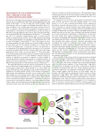
effects of luminal nutrients on the intestinal immune components. 27-29 CD T cells (Th0). The specific cytokines that are generated ultimately
4
The intestinal microbiota and the function and structure of the GI affect the differentiation pathway of these lymphocytes (Fig. 20-1).
32
tract are altered by changes brought on by critical illness. In the setting With gut disuse and fasting in critical illness, contractility is decreased,
levels within the levels) suppresses growth of
of increasing oxidative stress, where the pH or P O 2 the hostile environment (low pH or P O 2
lumen of the gut may drop, pathogenic bacteria like Pseudomonas and commensal organisms, and overgrowth of pathogenic bacteria occurs.
staphylococci undergo quorum sensing. If the number of organisms is These changes together with the absence of food antigen cause the
high enough, these pathogenic bacteria express virulent genes, which dendritic cell (which has been sampling the luminal contents) to release
allows adherence to the intestinal surface and a contact-dependent acti- interleukin-12 (Fig. 20-2). This cytokine causes naïve CD4 helper
vation of the intestinal epithelial cell. A cytokine storm results with the lymphocytes within the lamina propria to differentiate into a Th1 pro-
release of inflammatory agents (interleukin-1, interleukin-8, and tumor inflammatory subset. This Th1 response results in the further release of
necrosis factor) into lymphatic channels. A gut-lung conduit of inflam- other inflammatory cytokines, such as interleukin-2 (IL-2), interferon-γ
mation results, as these cytokines pass through lymphatic channels and (IFN-γ), and tumor necrosis factor-α (TNF-α). Feeding supports the
mesenteric lymph nodes into the thoracic duct and ultimately into the presence and role of commensal bacteria. In a fed state with food antigen
IL-2, IL-5
IL-12 IF-
+ TNF-
−
Innate immune system Th0 Inflamatory IFN- IL-10 Cellular
−
Down
Regulation
+ IL-4
IL-5
IL-6
IL-4
IL-10
? Tolerance IL-13
Microbes
Dendritic Humoral
Macrophage Tr1 IL-10 TGF-
IF-
FIGURE 20-1. Antigen processing immune function by the gut.
section02.indd 133 1/13/2015 2:04:54 PM