
Page 318 - Hall et al (2015) Principles of Critical Care-McGraw-Hill
P. 318
222 PART 2: General Management of the Patient
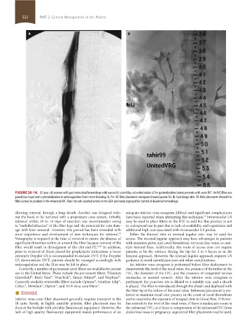
FIGURE 30-14. 62-year-old woman with gastrointestinal hemorrhage with successful control by coil embolization of the gastroduodenal artery presents with acute DVT. An IVC filter was
placed due to patient’s contraindication to anticoagulation from recent bleeding. A. Pre-IVC filter placement venogram showed patent IVC. B. Spot image after IVC filter placement showed the
filter (arrow) in position in the infrarenal IVC. Note the coils (dashed arrow) in the GDA previously deployed for control of duodenal hemorrhage.
allowing removal through a long sheath. Another was designed with- adequate inferior vena cavagram difficult and significant complications
out the hook to be retrieved with a proprietary cone system. Initially, have been reported when attempting this technique. Intravascular US
55
retrieval within 10 to 14 days of insertion was recommended owing may be used to place filters in the ICU as well but this practice is not
to “endothelialization” of the filter legs and the potential for vein dam- in widespread use in part due to lack of availability and experience and
age with later removal. However, this period has been extended with additional high cost associated with intravascular US probes.
more experience and development of new techniques for retrieval. Either the femoral vein or internal jugular vein may be used for
53
Venography is required at the time of retrieval to ensure the absence of access. The internal jugular approach may have advantages in patients
significant thrombus within or around the filter because removal of the with extensive pelvic and caval thrombosis, tortuous iliac veins, or mul-
filter would result in dislodgment of this clot and PE. 54,55 In addition, tiple femoral lines. Additionally, this route of access does not require
prior to retrieval of filters placed for prophylactic indications, a lower patients to lie flat without flexing the hip for 2 to 4 hours as in the
extremity Doppler US is recommended to exclude DVT. If the Doppler femoral approach. However, the internal jugular approach requires US
US demonstrates DVT, patients should be managed accordingly with guidance to avoid carotid puncture and other complications.
anticoagulation and the filter may be left in place. An inferior vena cavagram is performed before filter deployment to
Currently, a number of permanent caval filters are available for patient demonstrate the level of the renal veins, the presence of thrombus in the
use in the United States. These include the permanent filters: Titanium IVC, the diameter of the IVC, and the presence of congenital venous
Greenfield™, Bird’s Nest™, VenaTech™, Simon Nitinol™, and TrapEase™. anomalies or normal variants. After the inferior vena cavagram is
Currently available retrievable filters include Optease™, Gunther tulip™, performed, the puncture site is dilated to a suitable size, and a sheath
Celect™, Meridian™, Option™, and ALN vena cava filters™. is placed. The filter is introduced through the sheath and deployed with
■ TECHNIQUE the filter tip at the inflow of the renal veins. Infrarenal placement is pre-
ferred to maintain renal vein patency in the event of caval thrombosis
Inferior vena caval filter placement generally requires transport to the and to maximize the exposure of trapped clots to blood flow. If throm-
IR suite. Rarely, in highly unstable patients, filter placement may be bus extends to the level of the renal veins, if there is inadequate room in
done at the bedside with portable fluoroscopy equipment. However, the the infrarenal IVC, or if there is compression of the infrarenal IVC from
lack of high-quality fluoroscopy equipment makes performance of an abdominal mass or pregnancy, suprarenal filter placement may be used.
section02.indd 222 1/13/2015 2:05:59 PM