
Page 320 - Hall et al (2015) Principles of Critical Care-McGraw-Hill
P. 320
224 PART 2: General Management of the Patient
■ TECHNIQUE again obtained to confirm the reduction of the portosystemic gradient,
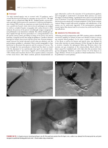
The right internal jugular vein is accessed with US guidance, and a and venography is performed to document shunt patency and assess
the degree of variceal filling. A gradient between portal vein and central
venous sheath is inserted from the venotomy site into the IVC. The right
hepatic vein is catheterized (Fig. 30-15). Wedged hepatic venous pres- veins of less than 12 mm Hg is desired because pressure gradients above
this level are associated with recurrent symptoms. If after shunt creation,
sures and right atrial pressures are measured to confirm portal hyperten-
sion. Using a TIPS needle set, punctures are made from the hepatic vein variceal filling is noted with portal venogram, coil embolization of the
varices can be performed, especially if the portosystemic pressures
into a portal vein branch. Some interventional radiologists use wedged
(or balloon occluded) hepatic venography with carbon dioxide to help remain higher than desired and the procedure was performed for bleed-
ing indications.
delineate the position of the portal vein although wedged venography is
fuses rapidly through the liver parenchyma in a retrograde fashion into ■ IMMEDIATE POSTPROCEDURE CARE
not performed at our institution routinely. The carbon dioxide gas dif-
the portal venous system, providing an image of the portal vein and its Following portal decompression with TIPS creation, patients should be
branches. Using this portal vein image for guidance, a needle is directed monitored carefully for at least 24 hours and should continue to receive
through the hepatic parenchyma into the portal vein. Pressure measure- any required blood products. In the past, a routine color duplex US
ments between the portal vein and central veins are obtained, and the of the shunt, as well as the hepatic and portal veins, was obtained 24
portosystemic gradient is calculated. Direct portal venography is then hours after insertion to assess adequacy of flow through the shunt and
performed to document the anatomy and the presence of varices. The to provide a baseline for subsequent follow-up. However, due to the
tract through the liver parenchyma is dilated and the shunt is created porosity and gas entrapment of the endoprosthesis, blood within the
with the placement of an endovascular stent-graft (or covered stent). shunt cannot be visualized until a few days after the procedure when
Currently, the Viatorr™ stent (Gore, Flagstaff, AZ) is widely preferred for gas has dissipated. Therefore, a routine 24-hour US of the shunt is no
TIPS creation and it has been shown to have superior patency compared longer obtained. If there is any question of shunt malfunction, CTA can
to bare metal stents. After shunt creation, pressure measurements are be performed to assess patency.
FIGURE 30-15. A. Hepatic venogram showed patent hepatic vein. B. After portal vein puncture from the hepatic vein, a catheter was advanced into the main portal vein, and portal
venogram was performed showing patent portal vein. C. Shunt creation using a covered stent.
section02.indd 224 1/13/2015 2:06:01 PM