
Page 566 - Hall et al (2015) Principles of Critical Care-McGraw-Hill
P. 566
386 PART 4: Pulmonary Disorders
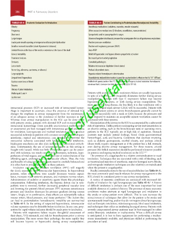
TABLE 45-4 Anatomic Evaluation for Intubation TABLE 45-5 Factors Contributing to Postintubation Hemodynamic Instability
Obesity Anesthesia medications (sedatives, narcotics, muscle relaxants)
Pregnancy Other vasoactive medications (β-blockers, vasodilators, vasoconstrictors)
Short neck Sympathetic and/or parasympathetic surges
Large tongue Absence of negative intrathoracic pressure that accompanies the loss of spontaneous respirations
Inadequate mouth opening or temporomandibular joint dysfunction Positive pressure ventilation
Small or recessed mandible (short thyromental distance) Positive end-expiratory pressure (PEEP)
Limited flexion at the base of the neck or extension at the base of the skull Auto-PEEP
https://kat.cr/user/tahir99/
Cervical instability Relief of hypercarbic and hypoxic driven sympathetic activation
Prominent incisors Decreased patient activity/agitation
Dentures Comorbid pathologies
Loose teeth Relative intravascular depletion (shock states)
Tumor (eg, adenoma, carcinoma, or abscess) Preload-dependent states
Large epiglottis Hypoxia-related hemodynamic deterioration
Lingual tonsil hyperplasia Hyperkalemia-induced deterioration (caused by succinylcholine’s effect on the Na /K ATPase)
+
+
Copious secretions or blood Modified with permission from Mort TC. Complications of emergency tracheal intubation: hemodynamic
Trauma alterations-Part I. J Intensive Care Med. May-June 2007;22(3):157-165.
History of prior intubations
Mallampati 3 and 4 Patients with acute hypoxemic respiratory failure are usually hypoxemic
Lip bite test in spite of a high Fi O 2 , and frequently desaturate further during airway
manipulation. Patients with type II respiratory failure may become
hypoxemic, hypercapneic, or both during airway manipulation. The
more severe the lung disease, the less likely it is that ventilation with a
intracranial pressure (ICP) or increased risk of intracranial hemor- mask or laryngeal mask airway (LMA) will be successful. Patients with
rhage is important to ascertain, since the presence of elevated ICP severe pulmonary edema or severe bronchospasm generally cannot be
changes the emphasis in airway management from the maintenance ventilated successfully with a mask or LMA because the pressures and
of an adequate airway to the avoidance of further increases in ICP. flows required to maintain an acceptable minute ventilation cannot be
Whereas most airway manipulation in the ICU can be done safely generated with these systems.
with patients awake, patients with elevated ICP and increased risk of Manipulation of the airway in the ICU is accompanied by a substantial
intracranial hemorrhage (from unstable arteriovenous malformations risk of aspiration. Unlike patients undergoing airway instrumentation in
or aneurysms) are best managed with intravenous general anesthesia an elective setting, such as the bronchoscopy suite or operating room,
for intubation. Laryngoscopy and tracheal intubation reliably produce patients in the ICU typically are at high risk of aspiration. Stomach
myocardial ischemia in patients with coronary artery disease. Adequate contents may include enteral feedings, blood (from gastrointestinal
anesthesia—topical and intravenous—can attenuate or prevent the hemorrhage), acid, and bacteria. Conditions that decrease emptying,
myocardial ischemia associated with laryngoscopy and intubation. such as diabetic gastroparesis, morbid obesity, and perhaps critical
Inadequate anesthesia can also elicit ischemia and associated arrhyth- illness itself, require management as if the patient has a full stomach,
mias. Unfortunately, the use of intravenous agents in this setting is even during elective airway management. For these reasons, cricoid
fraught with hazard. While too little intravenous agent can be associ- pressure (the Sellick maneuver) should be performed whenever possible
ated with ischemia, too much can cause hypotension, ischemia, hypo- on patients undergoing tracheal intubation in the ICU. 1,3-5
perfusion of vital organs, and a decreased rate of redistribution of the The presence of a coagulopathy is a relative contraindication to nasal
offending agent, prolonging its cardiovascular effects. Thus, the risks intubation. Techniques that are associated with a risk of bleeding, such
and benefits of using intravenous agents must be carefully balanced and as transtracheal injection of anesthesia, superior laryngeal nerve blocks,
it is often best to avoid using them in these patients. and retrograde intubation techniques are also relatively contraindicated
Intubation and positive pressure ventilation (PPV) will magnify when the patient is coagulopathic.
the shock associated with intravascular hypovolemia. In hypovolemic Finally, contraindications to the use of succinylcholine (see Table 45-3),
patients, reflex sympathetic tone usually decreases venous capaci- the most commonly used muscle relaxant for airway management in the
tance, increases mean systemic pressure, and maintains venous return. ICU, should be considered prior to any airway manipulation.
Administration of sedative or anesthetic agents blunts this physiologic A variety of anatomic conditions are associated with increased dif-
compensation. Following intubation, the hypoxic driven rise in sym- ficulty of intubation by rigid laryngoscopy (see Table 45-4). A history
6
pathetic tone is removed, further decreasing peripheral vascular tone of difficult intubation is perhaps one of the most important but least
and lowering the patient’s blood pressure. PPV increases intrathoracic available elements of a patient’s history. The presence of many anatomic
pressure and therefore decreases the pressure gradient driving venous conditions makes attempts at rigid laryngoscopy and intubation in
return. Singly, or in combination, these effects can substantially reduce the awake or asleep patient more difficult. This in turn increases the
venous return, blood pressure, and tissue perfusion. The factors that attractiveness of techniques that allow for the patient to be awake and
2
can lead to postintubation hemodynamic instability are summarized spontaneously breathing, and/or that do not require direct laryngoscopy,
in Table 45-5. In the setting of suspected hypovolemia, intravascular such as fiberoptic intubation, videolaryngoscopy, blind nasal intubation,
volume expansion may be desirable before intubation. In any case, prep- and techniques that utilize an intubating laryngeal airway. Patients with
aration for rapid volume infusion should be made prior to intubation. severely compromised airway anatomy may be best managed by either
Patients with respiratory failure require thoughtful assessment of awake fiberoptic intubation or tracheostomy. When a difficult airway
their shunt, V/Q mismatch, and risk for bronchospasm prior to airway is anticipated, it is best to have equipment for performing a tracheo-
manipulation. The more severe their pathology, the more rapidly they stomy immediately available, and physicians skilled at performing the
will become hypoxic or hypercarbic during airway manipulation. procedure at hand.
section04.indd 386 1/23/2015 2:18:46 PM