
Page 584 - Hall et al (2015) Principles of Critical Care-McGraw-Hill
P. 584
404 PART 4: Pulmonary Disorders
CHAPTER Upper Airway Obstruction
• Helium-oxygen mixtures reduce the density-dependent pressure
47 Brian K. Gehlbach required to drive airflow across obstructing upper airway lesions,
and may stabilize patients with UAO pending definitive therapy.
John P. Kress
• Prompt evaluation and management of suspected UAO may pre-
vent subsequent complications including cardiac arrest, anoxic
brain injury, and negative pressure pulmonary edema.
KEY POINTS
• Suspected upper airway obstruction (UAO) constitutes a medical
emergency. The immediate bedside consultation of a clinician
experienced in the management of this condition is indicated. There are few medical conditions that are as rapidly and predictably
• The initial evaluation of UAO is focused on determining the sever- lethal as the loss of upper airway patency. Because of the relative infre-
ity and suspected site of the obstruction. Arterial desaturation is quency with which upper airway obstruction (UAO) is encountered by
a late manifestation; better indicators of severity include stridor, most physicians, opportunities to acquire significant clinical experience
poor air movement, accessory muscle use, abnormal mentation or are limited. This, combined with the frequently subtle presentation of
agitation, tachycardia, hypertension, and pulsus paradoxus. upper airway obstruction and the clinician’s inability to visualize the
• Infections represent important causes of oropharyngeal and hypo- upper airway in its entire extent through routine physical examina-
pharyngeal UAO and include Ludwig angina, peritonsillar abscess, tion, may hamper diagnosis of this condition until a crisis results.
and infections of the retropharyngeal and lateral pharyngeal This chapter describes an approach to diagnosing and treating UAO
spaces. Otolaryngology consultation is indicated. Depending on as it presents in adults. While certain infections of the head, neck, and
the initial site of infection, spread to other critical sites (eg, the upper respiratory tract are considered here as they relate to UAO, the
mediastinum) may occur. specific approach to their management is considered in greater detail in
• While intubation is not always required in adults with epiglottitis, Chap. 73. A high index of suspicion for UAO, combined with early
consultation of anesthesia and otolaryngology services, is critical to the
management in an ICU is mandatory, and intubation equipment successful management of this condition.
and a tracheostomy tray should be at the bedside.
• Bacterial infections of the larynx are life-threatening. Causative ANATOMY OF THE UPPER AIRWAY
organisms include Staphylococcus aureus, Streptococcus pneu-
moniae, Haemophilus influenzae, and Corynebacterium diphtheriae. The upper airway comprises air-conducting passages that begin at
1,2
• Laryngospasm and laryngeal edema are important causes of the mouth or nose and end at the mainstem carina. The thoracic
postextubation stridor. Prophylactic corticosteroids may be inlet divides the upper airway into the intrathoracic and extrathoracic
airways. The extrathoracic airways are further divided into the naso-
effective at preventing this phenomenon in high-risk patients.
A reasonable approach is to administer methylprednisolone pharynx, oropharynx, hypopharynx, larynx, and extrathoracic trachea.
Air inspired through the nose passes through the nasal cavities and
20 mg IV q4h beginning 12 to 24 hours prior to planned extu-
bation and continued until the tube is removed. Patients with enters the nasopharynx after exiting the nose by way of the posterior
nares. Airflow proceeds inferiorly through the nasopharynx, passes
postextubation stridor from laryngeal edema may be treated
with a short (eg, 24 hours) course of corticosteroids. posterior to the soft palate, and enters the oropharynx. Closure of the
soft palate allows inspiration of air through the mouth. Air passes infe-
• Long-term intubation may result in a variety of problems related riorly through the oropharynx to the hypopharynx, which begins just
to the upper airway, including endotracheal tube obstruction from superior to the hyoid bone, and passes the epiglottis, thereby entering
secretions, vocal cord injury, subglottic stenosis, and tracheal stenosis. the larynx.
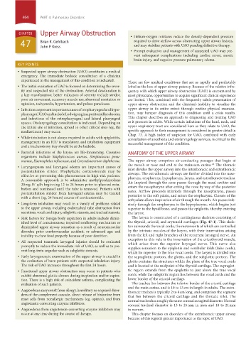
• Risk factors for foreign body aspiration in adults include dimin- The larynx is constructed of a cartilaginous skeleton consisting of
ished level of consciousness; impaired swallowing mechanism or the thyroid, cricoid, and arytenoid cartilages (Fig. 47-1). This skele-
diminished upper airway sensation as a result of neuromuscular ton surrounds the vocal cords, the movements of which are controlled
disorder, prior cerebrovascular accident, or advanced age; and by the intrinsic muscles of the larynx, with their innervation arising
inability to chew food properly because of poor dentition. from the left and right branches of the recurrent laryngeal nerve. An
• All suspected traumatic laryngeal injuries should be evaluated exception to this rule is the innervation of the cricothyroid muscle,
which arises from the superior laryngeal nerve. This nerve also
promptly to reduce the immediate risk of UAO, as well as to pre- supplies sensation to the epiglottis and vestibular folds (false cords),
vent long-term sequelae such as subglottic stenosis.
which lie superior to the true vocal cords. The larynx is divided into
• Early laryngoscopic examination of the upper airway is crucial in the supraglottic portion, the glottis, and the subglottic portion. The
the evaluation of burn patients with suspected inhalation injury. glottis contains the structures within the plane of the true vocal cords
The risk of UAO increases throughout the first 24 hours. and is located at the midpoint of the thyroid cartilage. The supraglot-
• Functional upper airway obstruction may occur in patients who tic region extends from the epiglottis to just above the true vocal
exhibit abnormal glottic closure during inspiration and/or expira- cords, while the subglottic region lies between the vocal cords and the
tion. There is a high risk of coincident asthma, complicating the lower border of the cricoid cartilage.
evaluation of such patients. The trachea lies between the inferior border of the cricoid cartilage
and the main carina, and is 10 to 13 cm in length in adults. The extra-
• Angioedema may result from allergy, hereditary or acquired disor- thoracic trachea is typically 2 to 4 cm long, and comprises the segment
ders of the complement cascade, direct release of histamine from that lies between the cricoid cartilage and the thoracic inlet. The
mast cells from nonallergic mechanisms (eg, opiates), and from normal trachea has roughly the same coronal as sagittal diameter. Normal
angiotensin-converting enzyme inhibitors. coronal tracheal diameter is 13 to 25 mm in men and 10 to 21 mm
• Angioedema from angiotensin-converting enzyme inhibitors may in women.
occur at any time during the course of therapy. This chapter focuses on disorders of the extrathoracic upper airway
because of this region’s greater importance to the topic of UAO.
section04.indd 404 1/23/2015 2:18:59 PM