
Page 696 - Hall et al (2015) Principles of Critical Care-McGraw-Hill
P. 696
CHAPTER 58: Restrictive Disease of the Respiratory System 515
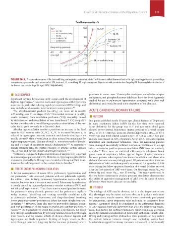
Total lung capacity – %
KEY 100 NL CW
NL CW
NL or scoliotic lung NL RS
NL RS 80
Scoliotic CW
Scoliotic RS Scoliotic CW
60 Scoliotic RS
Respiratory pressure
–40 –30 –20 –10 10 20 30 40
cms H O 40
2
NL or scoliotic lung
20
0
FIGURE 58-3. Pressure-volume curves of the chest wall, lung, and respiratory system in scoliosis. The P-V curve is shifted downward and to the right, requiring patients to generate large
transpulmonary pressures for small amounts of air. CW, chest wall; NL, normal lung; RS, respiratory system. (Reproduced with permission from Bergofsky EH. Respiratory failure in disorders of
the thoracic cage. Am Rev Respir Dis. April 1979;119(4):643-669.)
■ GAS EXCHANGE pressures in some cases. Prostacyclin analogues, endothelin receptor
1
Significant daytime hypoxemia rarely occurs until the development of antagonists, and phosphodiesterase inhibitors have not been rigorously
daytime hypercapnia. However, nocturnal hypercapnia with hypoxemia studied for use in pulmonary hypertension associated with chest wall
1
occurs early, particularly during rapid eye movement (REM) sleep, and deformities and should be used at the discretion of the clinician.
may underlie cardiovascular deterioration in some patients. 8,9 ACUTE CARDIOPULMONARY FAILURE
] on room air is usually
results primarily from ventilation-perfusion (V ˙ /Q ˙ ) inequality caused ■ OUTCOME
The alveolar-arterial gradient [(a-a)D O 2
1
≤25 mm Hg, even in late stages of KS. This modest increase in (a-a)D O 2
by atelectasis or underventilation of one hemithorax. V ˙ /Q ˙ inequality In a paper published nearly 30 years ago, clinical features of 20 patients
32
further contributes to a low diffusing capacity as does failure of the vas- in acute respiratory failure (ARF) for the first time were reported.
cular bed to grow normally in a distorted chest. Mean deformity for the group was 113° and admission blood gases
Alveolar hypoventilation results in part from an increase in the dead showed severe arterial hypoxemia (partial pressure of arterial oxygen
space to tidal volume ratio (V /V ). V /V is increased because V is [Pa O 2 ] of 35 ± 7 mm Hg), acute-on-chronic hypercapnia (Pa CO 2 of 63 ±
t
ds
t
ds
t
reduced in hypercapnic patients; anatomic and alveolar dead space are 9 mm Hg), and mild arterial acidemia (pH of 7.34 ± 0.08). Cor pul-
40
usually normal. Minute ventilation is often normal but maintained by monale was present in 60% of patients. Seven (35%) patients required
1
higher respiratory rates. The use of small V minimizes work of breath- intubation and mechanical ventilation, while the remaining patients
t
ing and is a sign of inspiratory muscle dysfunction. 30,33 As inspiratory were managed successfully without mechanical ventilation in an age
muscle strength falls, the partial pressure of arterial carbon dioxide when noninvasive positive-pressure ventilation (NIV) was not routinely
) rises and further impairs diaphragm function. 29,34 available. There were no statistical differences in admission blood
36
(Pa CO 2
Ventilatory response to high concentrations of inspired CO is normal gases, cause of respiratory failure, age, or degree of spinal curvature
2
in normocapnic patients with KS. However, in hypercapnic patients the between patients who required mechanical ventilation and those who
response is blunted by buffering from elevated cerebrospinal fluid bicar- did not. Outcome was surprisingly good. All patients survived their ini-
bonate or by a derangement in the central drive to breathe. 33 tial episode of ARF and subsequently experienced 2.4 episodes of ARF
■ EFFECTS ON THE PULMONARY CIRCULATION each during the follow-up period (median of 6 years). Median survival
was
was 55 mm Hg. This study performed in
A further consequence of severe KS is pulmonary hypertension and after the first episode of ARF was 9 years. On discharge, mean Pa O 2
63 mm Hg and mean Pa CO 2
cor pulmonale. Left untreated, patients with cor pulmonale typically the era before noninvasive positive pressure ventilation demonstrates
7
die within 1 year. Initially, pulmonary hypertension occurs only with the utility of aggressive management of ARF, even in cases of severe
3
exercise, but over time it occurs at rest as well. Pulmonary hypertension deformity. More recent data are not available.
not left atrial hypertension. Thus, there is an increased gradient between ■ ETIOLOGY
is usually caused by increased pulmonary vascular resistance (PVR) and
1
the pulmonary artery diastolic pressure and the pulmonary capillary The etiology of ARF may be obvious, but it is also important to note
wedge pressure. Identifying and treating reversible conditions such that the trigger may be minor and even obscure in patients with mini-
as pulmonary embolism, hypoxemia, and sleep-disordered breathing mal respiratory muscle reserve. ARF is most commonly precipitated
lowers pulmonary artery pressure and delays the onset of right ventricu- by pneumonia, upper respiratory tract infection, or congestive heart
lar failure. 36-39 However, there also may be irreversible changes associ- failure. Aspiration should be considered in the differential diagnosis
40
ated with proliferation of the media in smaller, pre-capillary pulmonary of ARF because chest wall deformity may affect the swallowing mecha-
vessels. The mechanism by which this occurs is not known, but blood nism. Risk factors for clotting (pulmonary hypertension and decreased
3,32
flow through vessels narrowed by low lung volumes, blood flow through mobility) mandate consideration of pulmonary embolism. Finally, iden-
fewer vessels, and the vascular effects of chronic alveolar hypoxia and tifying and treating airflow obstruction when possible can help restore
hypercapnia are likely important. Kinking of larger vessels as they the delicate balance between strength and respiratory system load.
travel through deformed lung may further increase pulmonary artery Limited data suggest that airway resistance is increased in mechanically
section04.indd 515 1/23/2015 2:20:28 PM