
Page 692 - Hall et al (2015) Principles of Critical Care-McGraw-Hill
P. 692
CHAPTER 57: Massive Hemoptysis 511
Small doses of codeine or morphine may be used to attenuate the < 35% of the time). CT scanning may be slightly more effective in local-
cough reflex to allow for clot formation. However, coughing is an effec- izing the site of bleeding, but is much better at determining the etiology
tive method to clear the airway, and a depressed sensorium may increase of the bleeding in part because bronchiectasis is much more evident.
8
the risk of aspiration. Therefore, these medications should be used with If urgent therapeutic stabilization is required, bronchoscopy is the pre-
discretion. ferred initial approach because the procedure also has the potential to
be therapeutic (eg, suctioning, endobronchial therapy, balloon tampon-
EVALUATION ade). When CT scan is performed, multidetector row CT scan should be
utilized as it is very effective at identifying bronchial, nonbronchial and
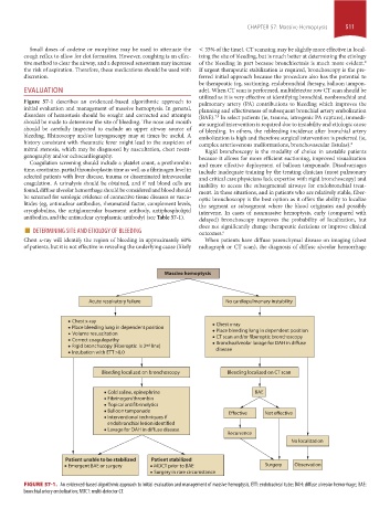
Figure 57-1 describes an evidenced-based algorithmic approach to pulmonary artery (PA) contributions to bleeding which improves the
initial evaluation and management of massive hemoptysis. In general, planning and effectiveness of subsequent bronchial artery embolization
disorders of hemostasis should be sought and corrected and attempts (BAE). In select patients (ie, trauma, iatrogenic PA rupture), immedi-
7,9
should be made to determine the site of bleeding. The nose and mouth ate surgical intervention is required due to instability and etiologic cause
should be carefully inspected to exclude an upper airway source of of bleeding. In others, the rebleeding incidence after bronchial artery
bleeding. Rhinoscopy and/or laryngoscopy may at times be useful. A embolization is high and therefore surgical intervention is preferred (ie,
history consistent with rheumatic fever might lead to the suspicion of complex arteriovenous malformations, bronchovascular fistulas). 6
mitral stenosis, which may be diagnosed by auscultation, chest roent- Rigid bronchoscopy is the modality of choice in unstable patients
genography and/or echocardiography. because it allows for more efficient suctioning, improved visualization
Coagulation screening should include a platelet count, a prothrombin and more effective deployment of balloon tamponade. Disadvantages
time, creatinine, partial thromboplastin time as well as a fibrinogen level in include inadequate training by the treating clinician (most pulmonary
selected patients with liver disease, trauma or disseminated intravascular and critical care physicians lack expertise with rigid bronchoscopy) and
coagulation. A urinalysis should be obtained, and if red blood cells are inability to access the subsegmental airways for endobronchial treat-
found, diffuse alveolar hemorrhage should be considered and blood should ment. In these situations, and in patients who are relatively stable, fiber-
be screened for serologic evidence of connective tissue diseases or vascu- optic bronchoscopy is the best option as it offers the ability to localize
litides (eg, antinuclear antibodies, rheumatoid factor, complement levels, the segment or subsegment where the blood originates and possibly
cryoglobulins, the antiglomerular basement antibody, antiphospholipid intervene. In cases of nonmassive hemoptysis, early (compared with
antibodies, and the antinuclear cytoplasmic antibody) (see Table 57-1). delayed) bronchoscopy improves the probability of localization, but
■ DETERMINING SITE AND ETIOLOGY OF BLEEDING does not significantly change therapeutic decisions or improve clinical
outcomes.
6
Chest x-ray will identify the region of bleeding in approximately 60% When patients have diffuse parenchymal disease on imaging (chest
of patients, but it is not effective in revealing the underlying cause (likely radiograph or CT scan), the diagnosis of diffuse alveolar hemorrhage
Massive hemoptysis
Acute respiratory failure No cardiopulmonary instability
Chest x-ray Chest x-ray
Place bleeding lung in dependent position
Volume resuscitation Place bleeding lung in dependent position
CT scan and/or fiberoptic bronchoscopy
Correct coagulopathy
Rigid bronchscopy (Fiberoptic is 2 nd line) Bronchoalveolar lavage for DAH in diffuse
disease
Intubation with ETT >8.0
Bleeding localized on bronchoscopy Bleeding localized on CT scan
Cold saline, epinephrine BAE
Fibrinogen/thrombin
Topical antifibrinolytics
Balloon tamponade Effective Not effective
Interventional techniques if
endobronchial lesion identified
Lavage for DAH in diffuse disease
Recurrence
No localization
Patient unable to be stabilized Patient stabilized
Emergent BAE or surgery MDCT prior to BAE Surgery Observation
Surgery in rare circumstance
FIGURE 57-1. An evidenced-based algorithmic approach to initial evaluation and management of massive hemoptysis. ETT: endotracheal tube; DAH: diffuse alveolar hemorrhage; BAE:
bronchial artery embolization; MDCT: multi-detector CT.
section04.indd 511 1/23/2015 2:20:24 PM