
Page 691 - Hall et al (2015) Principles of Critical Care-McGraw-Hill
P. 691
510 PART 4: Pulmonary Disorders
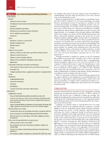
TABLE 57-1 Causes of Massive Hemoptysis and Pulmonary Hemorrhage For example, tuberculosis is the most common cause of hemoptysis in
underdeveloped countries while bronchiectasis is the most common
Localized Bleeding cause in the industrialized world. 1-2
Infections Massive hemoptysis has been variably defined as production of more
Mycobacteria (TB most common) than 300 to 600 mL of blood in 12 to 24 hours, depending on the study.
However, estimating the amount of hemoptysis is unreliable and has
Necrotizing bacterial pneumonia (particularly Klebsiella and Staphylococcus aureus)
very little clinical utility. Accordingly, a “magnitude-of-effect” is the pre-
Lung abscess ferred clinical approach as it uses the clinical consequences of hemop-
3
Mycetoma (Aspergillus most common) tysis such as airway obstruction, hypoxemia, hemodynamic instability
and underlying cardiopulmonary abnormalities to guide treatment and
Bronchiectasis (eg, cystic fibrosis or immune deficiencies)
triage decisions. As an example of this principle, patients with diffuse
Parasites (Hydatid cyst, paragonimiasis) alveolar hemorrhage can present with life-threatening hypoxemia and
Leptospirosis diffuse parenchymal infiltrates, meeting all the criteria for the acute
Tumors respiratory distress syndrome (ARDS), yet have little or no hemoptysis.
Bronchogenic carcinoma (ie, squamous cell) The conditions listed in Table 57-1 most often associated with massive
hemoptysis are: bronchiectasis, mycetoma, tuberculosis, bronchogenic
Pulmonary metastatic disease carcinoma, necrotizing pneumonia, and vascular-bronchial fistulas. The
Bronchial adenoma relative incidence of these conditions depends on the origin of the case
series examined. For example, when examining recent case series from
Sarcoma
China, Singapore, France, and Austria the relative prevalence of each
Pulmonary vascular problems condition differed: bronchiectasis (23%, 66%, 40%, 9%), tuberculosis
Pulmonary arteriovenous malformations (eg, Rendu-Osler-Weber syndrome) (55%, 10%, 14%, 23%), bronchogenic carcinoma (6%, 7%, 17%, 35%),
Pulmonary embolus with infarction respectively. 1,3-5
Although hemoptysis is classified as massive in only 5% of patients
Pulmonary aneurysm (eg, Behçet syndrome)
this condition is associated with a mortality of 9% to 38%, with death
4,6
Pulmonary artery catheterization with pulmonary arterial rupture attributed to asphyxiation more commonly than to exsanguination.
Mitral stenosis Massive hemoptysis originates from the bronchial circulation in 90%
of patients. Nonbronchial systemic vessels (5%) and pulmonary vessels
7
Coagulopathy (usually requires coexisting mucosal disruption)
(5%) are rarely the primary source of bleeding (although they make
Thrombocytopenia or platelet dysfunction (eg, von Willebrand disease, uremia) some contribution to the bleeding in up to a third of cases). Bleeding
7
Hemophilia A or B from vessels other than bronchial arteries most commonly occurs as a
result of inflammatory or infectious lung diseases causing anastomoses
Prolonged coagulation tests (due to coagulation factor production or consumption defect)
and collateral vessels to develop at the site of injury. 6
Trauma While it is important to determine whether bleeding originates from
Miscellaneous the lungs as opposed to the nasopharynx or gastrointestinal tract, the
Lymphangioleiomyomatosis distinction can generally be made by performing a careful history and
physical examination. In addition, blood coming from the lungs is usu-
Catamenial (endometriosis) ally bright red and has an alkaline pH, whereas that from the stomach
Cryptogenic is dark and acidic.
Broncholithiasis
Sarcoidosis (usually from cavitary lesions with mycetoma) STABILIZATION
Diffuse Bleeding Attention should initially be directed at airway management, ensuring
Capillaritis (seen on biopsy) adequate gas exchange and systemic perfusion. As little as 400 mL of
blood in the alveolar space is sufficient to impair gas exchange. The
Drug- and chemical-induced (propylthiouracil, phenytoin, retinoic acid)
likelihood of asphyxia from aspirated blood can probably be reduced by
Connective tissue diseases (ie, systemic lupus erythematosus, rheumatoid arthritis, localizing the site of bleeding and placing it in the dependent position
mixed connective tissue disease, systemic sclerosis, antiphospholipid antibody syn- (eg, left lateral decubitus at 45° reverse Trendelenburg if the site of bleed-
drome, polymyositis) ing is in the left lower lobe).
Systemic vasculitides (Behçet, cryoglobulinemia, IgA nephropathy, microscopic poly- Coughing is an effective means of clearing blood from the airway
angiitis, granulomatous vasculitis, pauci-immune, Henoch-Schonlein purpura) and patients should not be intubated unless gas exchange is critically
Bland hemorrhage (seen on biopsy) impaired or the rate of bleeding requires urgent therapeutic broncho-
scopic therapy (see below). In these situations, rigid bronchoscopy,
Connective tissue diseases (ie, systemic lupus erythematosus, Goodpasture syndrome) suctioning, and balloon tamponade should be performed prior to
Drugs (anticoagulant and antiplatelet therapy-glycoprotein IIa/IIIb inhibitors) intubation. Intubation with a large endotracheal tube (≥8) is preferred
6
Other (pulmonary veno-occlusive disease, mitral stenosis, idiopathic pulmonary to prevent tube obstruction and to allow for more effective suctioning.
hemosiderosis) Double lumen endotracheal tubes are generally not recommended, as
placing them is associated with substantial risks in the rapidly bleeding
Diffuse alveolar damage with bleeding (seen only on biopsy)
patient. In extreme circumstances, when bleeding is localized to the
6
Infection (any infection that can cause ARDS) right lung, the main stem bronchus of the left lung can be intubated.
Drugs (amiodarone, crack cocaine, nitrofurantoin, penicillamine, sirolimus, most This technique is not recommended when bleeding originates from the
cytotoxic drugs) left lung due to frequent occlusion of the right upper lobe by the endo-
Connective tissue diseases (systemic lupus erythematosus, rheumatoid arthritis, tracheal tube. An alternative and more effective means to manage local-
polymyositis) ized bleeding is to use a bronchoscopic-guided Fogarty balloon catheter
(larger airways) or a pulmonary artery catheter (distal subsegmental air-
Other (pulmonary capillary hemangiomatosis, pulmonary infarct, ARDS of any cause)
ways) to occlude the bleeding airway and protect the contralateral lung. 6
section04.indd 510 1/23/2015 2:20:23 PM