
Page 703 - Hall et al (2015) Principles of Critical Care-McGraw-Hill
P. 703
522 PART 4: Pulmonary Disorders
considerable financial burden imposed on the health care system by the episodes of VAP, only three variables remained significant: duration of
development of VAP. 26,48,49,51-55 mechanical ventilation of longer than 7 days before onset of VAP, prior
■ ETIOLOGIC AGENTS antibiotic use, and prior use of broad-spectrum drugs (third-generation
cephalosporins, fluoroquinolones and/or imipenem). Not all studies
59
Microorganisms responsible for VAP differ according to the population have confirmed this distribution pattern, and in some studies the most
of ICU patients, the durations of hospital and ICU stays, and the spe- common pathogens associated with early-onset VAP were P. aeruginosa,
cific diagnostic method(s) used to establish the responsible pathogens. MRSA and Enterobacter spp, with similar pathogens associated with
A number of studies have shown that gram-negative bacilli (GNB) cause late-onset VAP. 60,61 These findings might be explained in part by prior
many of the respiratory infections in this setting. 1,3,56,57 The data from 24 hospitalization and the use of antibiotics before transfer to the ICU.
studies conducted on ventilated patients, for whom bacteriologic studies The incidence of multiresistant pathogens is also closely linked
were restricted to uncontaminated specimens obtained using a protected to local factors and varies widely from one institution to another.
specimen brush (PSB) or bronchoalveolar lavage (BAL), confirmed these Consequently, each ICU has to continuously collect meticulous epide-
62
results: GNB represented 58% of recovered organisms (Table 59-1). The miologic data. Clinicians clearly must be aware of the common micro-
3
predominant GNB were P. aeruginosa and Acinetobacter spp, followed organisms associated with both early-onset and late-onset VAP in their
by Proteus spp, Escherichia coli, Klebsiella spp, and H. influenzae. A rela- own hospitals in order to avoid the administration of initial inadequate
tively high rate of gram-positive pneumonias was also reported in those antimicrobial therapy.
studies, with S. aureus involved in >20% of the cases. Many episodes of Legionella species, anaerobes, and even Pneumocystis jirovecii should
56
VAP are caused by multiple pathogens. 3,58 be mentioned as potential causative agents, but these microbes are
Underlying diseases may predispose patients to infection with spe- not commonly found when pneumonia is acquired during mechani-
cific organisms. Patients with chronic obstructive pulmonary disease cal ventilation. Herpesviridae, namely herpes simplex virus (HSV)
(COPD) are at increased risk for H. influenzae, Moraxella catarrhalis, or can be detected in the lower respiratory tracts of 5% to 64% of ICU
S. pneumoniae infections; cystic fibrosis increases the risk of P. aerugi- patients, depending on the population and the diagnostic method
nosa and/or S. aureus infections, while trauma and neurological disease used. In most cases, HSV recovery from lower respiratory tract samples
increases the risk for S. aureus infection. Furthermore, the causative of nonimmunocompromised ventilated patients corresponds to viral
agent for pneumonia differs among ICU surgical populations, with 18% contamination from the mouth and/or throat. For some patients, how-
of the nosocomial pneumonias caused by Haemophilus or pneumococci, ever, real HSV bronchopneumonitis can develop and it can evolve into
particularly in patients with trauma, but not in patients with malig- ARDS and/or facilitate the occurrence of bacterial superinfection. 63-65
nancy, transplantation, abdominal or cardiovascular surgery. 1,3 Cytomegalovirus-induced pneumonia is a rare event in ventilated
Despite somewhat different definitions of early-onset pneumonia, patients. As for HSV bronchopneumonitis, it is impossible to know
varying from onset of less than 3 to less than 7 days, high rates of whether CMV detection in the lower respiratory tract is merely a marker
H. influenzae, S. pneumoniae, MSSA or susceptible Enterobacteriaceae of disease severity or signals real disease with its own morbidity and
were constantly found in early-onset VAP, whereas P. aeruginosa, mortality. 66-69
Acinetobacter spp, MRSA and multiresistant GNB were significantly Isolation of fungi, most frequently Candida species, at significant
more frequent in late-onset VAP. The different pattern of distribution concentrations poses interpretative problems. Invasive disease has been
3
of etiologic agents between early- and late-onset VAP is linked to prior reported in VAP but yeasts are isolated more frequently from respiratory
antimicrobial therapy in many patients with late-onset VAP. When tract specimens in the absence of apparent disease, even when retrieved
multivariate analysis was used to identify risk factors for VAP caused at high concentrations from bronchoscopic specimens. 70-74 Thus, based
by potentially drug-resistant bacteria such as MRSA, P. aeruginosa, on current data, the presence of yeasts in respiratory secretions obtained
Acinetobacter baumannii, and/or S. maltophilia in 135 consecutive from non-immunosuppressed ventilated patients usually indicates
colonization rather than infection of the respiratory tract, and does not
justify by itself a specific antifungal therapy. Evidence of lung tissue
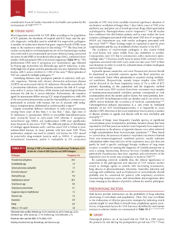
TABLE 59-1 Etiology of VAP as Documented by Bronchoscopic Techniques in 24 invasion is needed for making the diagnosis of Candida pneumonia in
Studies for a Total of 1689 Episodes and 2490 Pathogens such a setting. Interactions, however, between Candida and bacteria,
particularly Pseudomonas, have been reported, and colonization of the
Pathogen Frequency (%) respiratory tract by yeasts may predispose to bacterial VAP. 75-78
Pseudomonas aeruginosa 24.4 By examining currently available data, the clinical significance of
anaerobes in the pathogenesis and outcome of VAP remains unclear
Acinetobacter spp 7.9
except as etiologic agents in patients with necrotizing pneumonitis,
Stenotrophomonas maltophilia 1.7 lung abscess or pleuropulmonary infections. Anaerobic infection and
Enterobacteriaceae a 14.1 coverage with antibiotics, such as clindamycin or metronidazole, should
Haemophilus spp 9.8 probably also be considered for patients with respiratory secretions
documenting numerous extra- and intracellular microorganisms after
Staphylococcus aureus b 20.4 Gram staining in the absence of positive cultures for aerobic pathogens.
Streptococcus spp 8.0
Streptococcus pneumoniae 4.1 PREDISPOSING FACTORS
Coagulase-negative staphylococci 1.4
Risk factors provide information on the probability of lung infection
Neisseria spp 2.6 developing in individuals and populations. Thus, they may contribute
Anaerobes 0.9 to the elaboration of effective preventive strategies by indicating which
Fungi 0.9 patients might be most likely to benefit from prophylaxis against pneu-
monia. Independent factors for VAP that were identified by multivariate
Others (<1% each) c 3.8
analyses in selected studies are summarized in Table 59-2. 2,11,13,14,29,51,79-83
a Distribution when specified: Klebsiella spp, 15.6%; Escherichia coli, 24.1%; Proteus spp, 22.3%;
Enterobacter spp, 18.8%; Serratia spp, 12.1%; Citrobacter spp, 5.0%; Hafnia alvei, 2.1%. ■ SURGERY
b Distribution when specified: MRSA, 55.7%; MSSA, 44.3%. Postsurgical patients are at increased risk for VAP. In a 1981 report,
84
c Including Corynebacterium spp, Moraxella spp, and Enterococcus spp. the pneumonia rate during the postoperative period was 17%. Those
section04.indd 522 1/23/2015 2:20:32 PM