
Page 822 - Hall et al (2015) Principles of Critical Care-McGraw-Hill
P. 822
CHAPTER 62: Sepsis and Immunoparalysis 553
EVOLUTION TO IMMUNOPARALYSIS (Fig. 62-1) 31 their corresponding ligands (eg, PD-1 ligand [PD-L1]). Furthermore,
the numbers of regulatory T cells and myeloid-derived suppressor
As described above, much of our therapy for sepsis is “early and goal cells (MDSCs) are increased, and there is a shift from a phenotype
directed” and unfolds in the early phase of the syndrome during which of inflammatory type 1 helper T (Th1) cells to an anti-inflammatory
large and uncontrolled release of endogenous and exogenous mediators phenotype of type 2 helper T (Th2) cells characterized by the production
of inflammation occurs. Despite our efforts to identify patients early and of interleukin-10.
deploy therapies such as early fluid therapy, mortality remains as high The net result is a severely compromised innate and adaptive
as 30% to 40% for the highest risk patients. 32-34 Much of this mortality immune system with poorly functional “exhausted” CD8 and anergic
occurs during the state of immunoparesis, related to “second and third CD4 T cells. Targets of potential immunotherapeutic approaches (see
hits” in the form of nosocomial superinfections. 34 Fig 62-1) include agents that block apoptosis, block negative costimu-
During this phase of sepsis activation of anti-inflammatory media- latory molecules, decrease the level of anti-inflammatory cytokines,
tors such as interleukin 10 (IL-10), and transforming growth factor- increase HLA-DR expression, and reactivate “exhausted” or anergic
beta (TGF-β) are well described, and it appears these pathways are T cells. FLT-3L denotes Fms-related tyrosine kinase 3 ligand, GM-CSF
activated in the course of strong proinflammatory stimulation, perhaps granulocyte– macrophage colony-stimulating factor, LFA-1 lymphocyte
as an effort to achieve homeostasis. Modification of cellular function is function–associated antigen 1, and tumor necrosis factor α (TNF-α).
another feature of this state of immunoparesis, and sustained periods of In this later, hypoimmune phase of the disease, hemodynamics will
monocyte deactivation characterized by defective phagocytosis, altered most likely be relatively stable. Mechanical ventilation is often neces-
antigen presentation, and reduced production of inflammatory cyto- sary, and a requirement for renal replacement therapy is common.
kines by these cells have been described. 25 The main focus of the treating clinician will now be on the prevention
This reprogramming event of immune function occurs at other levels and treatment of secondary infections, such as ventilator-associated
as well. At the transcriptional level, downregulation of genes encoding pneumonia, catheter-related infections, and fungal infections. The
proinflammatory cytokines and other acute phase proteins is accom- patient will most likely be treated with various antibiotic and antifun-
panied by an outpouring of anti-inflammatory cytokines and cytokine gal regimes, and multiresistant strains may be detected in the further
inhibitors, and downregulation of cytokine receptors. Neuroendocrine course of the disease.
35
response also appears to attenuate inflammatory cytokine synthesis and
In addition, specific subsets of lymphocytes, dendritic cells, and ■
also results in reduction of antigen expression on antigen presenting
cells. 25,36 DEFINITION OF SYSTEMIC SEPSIS-INDUCED IMMUNOPARALYSIS
epithelial cells undergo apoptosis at extremely accelerated rates after a In response to stress and injury (of which septic shock is a typical example),
typical septic stimulus in patients. 37,38 Uptake of apoptotic cells further the body develops compensatory mechanisms to prevent systemic
impairs host immunity by inducing an anti-inflammatory phenotype in inflammation. This anti-inflammatory response is a homeostatic mech-
phagocytic cells that consume the cellular corpses. Prevention of this anism that occurs in all patients. It protects against lethal overwhelm-
39
sepsis-induced apoptosis apparently attenuates the immunosuppressive ing inflammation during the first hours of the syndrome, but becomes
cascade and leads to sustained immunity. 37 deleterious as it persists because nearly all immune functions are com-
T-regulatory cells and myeloid-derived suppressor cells are also acti- promised. As all cell types—neutrophils, monocytes/macrophages, and
vated during the later phases of sepsis and appear to participate in general lymphocytes—are impaired, both innate and specific immunities are
downregulation of immune response and the inflammatory state. 39-42 depressed. The terms immunoparalysis and immunosuppression have
Other described mechanisms of this immunosuppression include been proposed to describe the global incapacity of the body to mount
massive apoptosis-induced depletion of lymphocytes and dendritic cells, any kind of immune response. 43
decreased expression of the cell-surface antigen–presenting complex In many cases of sepsis, the immune system fails to eradicate the
HLA-DR, and increased expression of the negative costimulatory mol- infectious pathogens, and a prolonged phase of sepsis-induced immu-
ecules programmed death 1 (PD-1), cytotoxic T-lymphocyte–associated nosuppression begins, characterized by a failure to eradicate the primary
antigen 4 (CTLA-4), and B- and T-lymphocyte attenuator (BTLA) and infection and by development of secondary nosocomial infections.
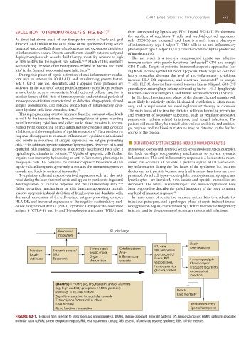
Recovery/ ICU discharge
resolution
Death
ICU care
Severe sepsis Cytokine storm Early mortality
Infection Systemic spread/ Septic shock Anti- MV, antibiotics
locally SIRS Organ inflammatory source control
at tissues Bacteremia Fluids, RRT Immunoparalysis
dysfunction cascade
vasopressors, Chronic sepsis
hemodynamics, Frequent/recurrent/
glucose control uncontrolled
infections
(DAMPs ) = PAMPs (eg, LPS, flagellin) and/or Alarmins
(eg, high-mobility group box 1 S100a proteins)
PRRs (eg, TLRs) cells surface Death
Late mortality
Signal transmission. Intracellular cascade
Transcription factors-cell nucleus
DNA binding Immune recovery
Gene function modulation (partial/complete)
FIGURE 62-1. Evolution from infection to septic shock and immunoparalysis. DAMPs, damage-associated molecular patterns; LPS, lipopolysaccharide; PAMPs, pathogen-associated
molecular patterns; PRRs, pattern recognition receptors; RRT, renal replacement therapy; SIRS, systemic inflammatory response syndrome; TLRs, Toll-like receptors.
section05_c61-73.indd 553 1/23/2015 12:47:15 PM