
Page 874 - Hall et al (2015) Principles of Critical Care-McGraw-Hill
P. 874
CHAPTER 68: Approach to Infection in Patients Receiving Cytotoxic Chemotherapy for Malignancy 605
reviews with meta-analyses have suggested an enhanced risk for serious that occurs during the second and third weeks. This stage is character-
grade 3 to 4 infectious complications associated with the use of rituximab ized histologically by cellular necrosis, lack of mitotic activity, and focal
for maintenance therapy in non-Hodgkin lymphoma patients. 87,88 Case loss of villous surfaces and clinically by abdominal pain, diarrhea, elec-
reports and small series have reported some opportunistic infections trolyte loss, and invasive infection. The third stage of cellular regenera-
associated with the use of rituximab, including tuberculosis, progressive tion occurs after the third week and is characterized by resumption of
multifocal leukoencephalopathy, babesiosis, pulmonary Pneumocystis mitotic activity and cellular proliferation in the crypts with subsequent
jirovecii infection, enteroviral gastroenteritis, cytomegalovirus infection, repopulation of the denuded surfaces by differentiated cells.
reactivation of hepatitis B virus infection, disseminated varicella-zoster, The maximum cytotoxic therapy–induced intestinal epithelial damage
and parvovirus B19–related pure red cell aplasia. occurs in the second week between days 10 and 14. 93-95 This corresponds
to the median time of onset of bacteremic infection on day 14 due to the
Integumental Barriers: Integumental barriers are among the most microorganisms that normally colonize these surfaces. To a limited
96
important and most often damaged defense systems for cancer extent, the type of pathogens recovered in bacteremic infections can
patients. These barriers include the epithelial surfaces of the skin, the be predicted from the pool of microorganisms colonizing damaged
upper and lower respiratory tract, the upper and lower gastrointestinal mucosal surfaces. Oral mucosal ulceration, particularly that involving
(GI) tract, and the mucosal surfaces lining the genitourinary tract. In periodontal tissues, is often associated with viridans group streptococcal
critically ill patients, the barrier function of these surfaces may also be bacteremia. 97,98 Colonic mucosal damage is more likely to be associated
compromised by procedures such as percutaneous intravenous cath- with aerobic gram-negative bacillary infection with Escherichia coli,
eterization, endotracheal intubation, endoscopic procedures, nasogas- Klebsiella species, Pseudomonas aeruginosa, or opportunistic yeasts
tric intubation, and indwelling urinary catheterization (Table 68-1). when these pathogens are colonizing the lower GI tract. 96
Integumental damage secondary to cytotoxic therapy has become Mucositis not only predisposes patients to invasive infection, but also
more prevalent as the dose intensity of the remission-induction regi- imposes a significant cost with respect to the resources needed to man-
mens has increased. The epithelial surfaces of the GI tract appear age the consequences of mucositis. 99,100 Recent cost estimates suggest
89
to be at greatest risk. The antiproliferative effect of therapy prevents that an episode of severe mucositis may cost an average of $7985 (Year
cell recruitment into mucosal areas denuded by erosion or by cellular 2002) per patient. Neutropenia with or without infection is estimated
100
attrition, resulting in the appearance of superficial erosion and ulcer- to cost an average of $9316 (Year 2002) per inpatient.
ation. The absorptive capacity of the GI mucosa may also be impaired
significantly among recipients of regimens such as HDARA-C, and both
anatomic mucosal disruption and absorptive dysfunction appear to INFECTIONS AND BACTERIAL PATHOGENS CAUSING
temporally parallel that of the neutrophil profile. NEUTROPENIC FEVERS
A high proportion of patients receiving cytoreductive therapy also
experience painful, often debilitating inflammatory lesions within A review of bloodstream infections occurring in patients with hemato-
the oral cavity. The tissues of the periodontium, gingival surfaces, logical malignancies over a 14-year period in a tertiary cancer center in
90
oral mucosa, and mucosal surfaces of the upper and lower bowel are Sweden noted that gram-negative bacilli accounted for 45% and gram-
101
affected. Cytotoxic regimens affect the developing basal epithelial cells positive organisms accounted for 55%. Of note in this experience
89
of the oral mucosa in a manner that parallels the effect on the marrow was the rising incidence of enterococcal bloodstream infections due to
system cell pool and the intestinal mucosal surface. Mucosal atrophy, penicillin-resistant E faecium and the high 30-day mortality (24%) com-
91
cytolysis, and denudation of the mucosal surface result in the painful pared to other gram-positive 30-day mortality rates (~15%). 101
foci of local ulceration typically observed 4 to 7 days after administra- In an Irish 5-year experience in febrile neutropenic cancer patients,
tion of cytotoxic agents, which usually resolve spontaneously between 20% of blood cultures revealed 172 isolates of which 123 (71%) were
days 14 and 21. 90,92 gram-positive organisms, 48 (28%) were gram-negative bacilli, and 2
102
Cytotoxic therapy–induced intestinal mucosal damage has been were yeasts. Of the gram-positive organisms, 93 were Staphylococcus
described in three stages. The first stage of initial injury begins during spp, 10 were Streptococcus spp, 11 were Enterococcus spp, and 9 were
91
the first week of cytotoxic therapy and is characterized by replacement predominantly gram-positive bacilli. The staphylococci were coagulase
of the normal crypts and mucus-secreting goblet cells by atypical undif- negative in 65 and S aureus in 28, of which 25 (89%) were methicillin
ferentiated cells. The second stage represents progressive mucosal injury resistant. This highlights the high incidence of methicillin resistance in
this population and has implications for the choice of initial empirical
antibacterial therapy.
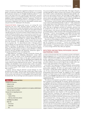
TABLE 68-1 Integumental Defects The infections documented among febrile neutropenic patients have
been classified as microbiologically documented with the identification
Damage to mucosal surfaces of a pathogen and a focus of infection; as clinically documented with the
Endotracheal tube identification of a clinical focus of infection without isolation of a putative
Nasogastric tube pathogen; and as an unexplained fever wherein neither a clinical focus nor
a pathogen are identified. Among febrile neutropenic cancer patients
103
Cytotoxic therapy–induced damage to gastrointestinal and respiratory epithelial barriers
not receiving fluoroquinolone chemoprophylaxis managed during the
Endoscopic diagnostic procedures early 1990s, Cornelissen and colleagues reported that microbiologically
Damage to skin and supporting structures documented infections were observed in 33% of patients with gram-
negative infections comprising 18%, gram-positive infections in 9%,
IV catheters
and mixed gram-negative and gram-positive in 6%. Forty-two percent
104
Peripheral IV lines of patients had clinically documented infections and the remaining 24%
Indwelling central venous catheters had unexplained fevers. Among a similar group of patients who had
received ciprofloxacin chemoprophylaxis, there were no gram-negative
Indwelling urinary catheters
infections. Gram-positive infections were observed in 38% of patients,
Biopsy sites clinically documented infections in 47% of patients, and unexplained
Bone marrow fevers in only 15%. Fluoroquinolone antibacterial chemoprophylaxis can
Lymph nodes reduce the risk for invasive gram-negative infections in patients at high-
risk for such infections in an environment where the prevalence of gram-
Skin
negative resistance to fluoroquinolone antibacterial agents is low. 21,105
section05_c61-73.indd 605 1/23/2015 12:48:04 PM