
Page 876 - Hall et al (2015) Principles of Critical Care-McGraw-Hill
P. 876
CHAPTER 68: Approach to Infection in Patients Receiving Cytotoxic Chemotherapy for Malignancy 607
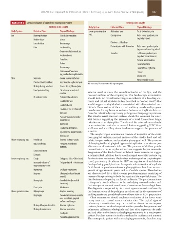
TABLE 68-2 Clinical Evaluation of the Febrile Neutropenic Patient Findings to Be Sought
Findings to Be Sought Body System Historical Clues Physical Findings
Body System Historical Clues Physical Findings Lower gastrointestinal Abdominal pain Focal abdominal pain
Eye Blurring of vision Scleral abnormalities tract Constipation Right upper quadrant pain
(eg, biliary tree)
Double vision Icterus
Loss of vision Hemorrhage Diarrhea ± bleeding
Pain Local swelling Perianal pain with defecation Right lower quadrant pain
(eg, cecum/ascending colon)
Conjunctival abnormalities
Jaundice Left lower quadrant pain
Focal erythema (eg, diverticular disease)
Petechiae Perianal abnormalities
Retina Focal tenderness
Hemorrhage Focal/diffuse erythema
“Cotton wool” exudates Fissures
(eg, candidal endophthalmitis)
Ulcerations
Skin Skin rash Central venous catheters
Hemorrhoidal tissues
Pruritus (focal or diffuse) Insertion site erythema/pain
HR, heart rate; IV, intravenous; RR, respiratory rate.
History of drug reactions Tunnel site erythema/pain
Focal pain/swelling Exit site erythema/pain/
exudate anterior nasal mucosa, the vermilion border of the lips, and the
mucosal surfaces of the oropharynx. The funduscopic examination
IV catheter site(s) Peripheral IV catheters should look for retinal hemorrhages as evidence of a bleeding dia-
Focal tenderness thesis and retinal exudates (often described as “cotton wool”) that
Focal erythema would suggest endophthalmitis associated with disseminated can-
didiasis. Examination of the external auditory canals and tympanic
Exudate at the insertion site membranes for erythema or vesicular lesions can implicate this as a
Skin rash focus for infection by respiratory pathogens or herpes group viruses.
Papular/macular/vesicular The anterior nasal mucosal surfaces should be examined for ulcer-
morphotypes ated lesions suggesting the presence of a local filamentous fungal
infection such as Aspergillus. The skin of the external nares should
Ulceration
be examined for vesicular or crusted lesions suggesting HSV. Nasal
Focal areas of necrosis stuffiness and maxillary sinus tenderness suggests the presence of
(eg, ecthyma gangrenosum) sinusitis.
The oropharyngeal examination consists of inspection of the denti-
Distribution
tion, gingival surfaces, mucosal surfaces of the cheeks, hard and soft
Upper respiratory tract Painful ear External auditory canals palate, tongue surfaces, and posterior pharyngeal wall. The presence
Nasal stuffiness Tympanic membrane of decaying teeth and gingival hyperemia implicates those sites as pos-
erythema sible sources of bacteremic infection. The presence of shallow, painful
mucosal ulcers on an erythematous base suggests herpes mucositis.
Sinus tenderness
Progression of this kind of lesion with local tissue necrosis can suggest
Epistaxis a polymicrobial infection due to oropharyngeal anaerobic bacteria (eg,
Lower respiratory tract Cough Tachypnea (RR >20/minute) Fusobacterium nucleatum, Bacteroides melaninogenicus, peptostrepto-
cocci), particularly if cultures for HSV are negative or if such lesions
Increased volume of Tachycardia (HR >90/minute) develop during prophylactic or therapeutic administration of acyclovir.
respiratory secretions
Oral thrush or pseudomembranous pharyngitis evolves from an over-
Hyperpnea Localized crepitations growth of opportunistic yeasts such as Candida species. These lesions
Dyspnea Effusions (reduced breath are characterized by a thick creamy pseudomembrane consisting of
sounds) masses of fungi existing in both the yeast and the mycelial phases. The
distribution may be patchy, confluent, or discrete. The pseudomembrane
Hemoptysis Consolidation (bronchial is frequently closely adherent to the underlying mucosal surface such
breathing)
that attempts at removal reveal an erythematous or hemorrhagic base.
Chest pain Friction rub The diagnosis is suspected by the clinical appearance and confirmed by
Upper gastrointestinal Odynophagia Gingival bleeding the demonstration of the pathogen in culture and by the appearance of
Dysphagia Pseudomembranous exudate budding yeasts and pseudohyphae on a Gram stain or KOH preparation.
over buccal and gingival Chest examination should emphasize evaluation of the lower respi-
surfaces and tongue ratory tract and central venous catheter sites. The typical signs of
pulmonary consolidation may be muted or absent in neutropenic
History of herpes stomatitis Mucosal erythema patients; however, localized crepitation often precedes the appearance of
History of denture use Mucosal ulceration pulmonary infiltrates radiologically and thus often represents the earli-
Focal pain est (and often only) clue to a developing pneumonia in a neutropenic
patient. Purulent sputum is similarly reduced in incidence and amount.
Preexisting periodontitis
The neutropenic patient with a developing pneumonia, therefore, may
section05_c61-73.indd 607 1/23/2015 12:48:05 PM