
Page 877 - Hall et al (2015) Principles of Critical Care-McGraw-Hill
P. 877
608 PART 5: Infectious Disorders
manifest only as febrile illness associated with an increased respiratory rashes associated with specific drugs such as β-lactam antibacterial
rate and a few localized crepitations, with or without an associated drugs, allopurinol, or trimethoprim-sulfamethoxazole (TMP/SMX); and
cough or radiologic changes. The clinician must search for additional specific chemotherapy regimen–related rash syndromes (eg, the exfo-
132
differential diagnostic clues such as the origin of the suspected pneu- liative palmar/plantar syndrome associated with high-dose cytarabine;
monia (community or hospital acquired), the tempo of the illness, the Fig. 68-4). These skin rash syndromes may coexist simultaneously.
association of the illness with other potentially noninfectious factors Once the relevant historical details and physical findings are established,
such as pulmonary edema, exposure to certain chemotherapeutic agents the complete evaluation of the febrile neutropenic patient should include a
associated with lung injury (bleomycin, busulfan, cytarabine), radiation series of laboratory and radiologic investigations designed to complement
therapy, pulmonary thromboemboli, pulmonary hemorrhage, or hyper- the clinical examination. Specimens of body fluids such as blood, urine,
leukocytosis. Chest physical examination can do little to differentiate cerebrospinal fluid, and lower respiratory secretions should be submitted to
infectious or noninfectious causes of pulmonary findings, but it can help the clinical microbiology laboratory for culture and antimicrobial suscep-
identify the lower respiratory tract as the potential infected focus. tibility testing where appropriate. At least two sets of blood cultures should
The symptoms and signs of an intra-abdominal infection may be be obtained, one of which should be from a peripheral venous site. Further,
obvious or muted, focal, or diffuse. The most important finding is it has been recommended that for patients with multilumen indwelling cen-
focal tenderness. For example, tenderness in the right lower quadrant tral venous catheters in situ, each lumen of the catheter should be sampled
130
might suggest neutropenic enterocolitis (typhlitis); right upper quadrant in addition to blood from the peripheral venous site. 120,129
tenderness, a biliary tract focus or hepatomegaly; epigastric pain, an The basic radiologic investigation is the chest radiograph. When
upper GI focus; and left lower quadrant tenderness, colitis or diverticu- suggested by clinical clues, sinus radiographs are useful for detecting
lar disease. It is important to examine the perianal tissues for signs of sinus opacification or fluid levels. Panorex radiographs can be help-
excoriation, local erythema, swelling, tenderness, fissure formation, or ful for evaluating periodontal infection. High-resolution computed
hemorrhoidal tissues, since this area is frequently the site of major life- tomographic (HRCT) examination of the lungs has a high yield of
threatening infection in neutropenic patients. Digital examination of abnormalities in febrile neutropenic patients despite nondiagnostic
the rectum is not recommended in neutropenic patients because of the chest radiographs. 133,134 In one study, 60% of febrile neutropenic patients
additional risk of tissue damage, bleeding, and infection. A light perianal with normal chest radiographs had a pulmonary infiltrate demonstrable
digital examination, however, can be informative about focal areas of on the chest HRCT. Computed tomography (CT) of the abdomen
133
cellulitis without increasing the risk of bacteremic infection. or hepatic ultrasonography is valuable for assessing the significance of
Examination of the skin should consist of a thorough search for abnormalities in cholestatic enzymes (γ-glutamyltransferase [GGT] and
focal areas of pain, swelling, or erythema, especially in association with alkaline phosphatase). This is particularly important if the possibility of
indwelling vascular access devices. Particular attention should be paid hepatosplenic candidiasis exists. Abdominal pain and tenderness with
to the venous insertion, tunnel, and exit sites associated with central diarrhea in a persistently febrile neutropenic patient suggests the possi-
venous catheters. In contrast, nonspecific local pocket tenderness may bility of neutropenic enterocolitis. Abdominal CT looking for bowel wall
be the only clue to infection associated with the totally implantable thickening, pneumatosis, wall nodularity, mucosal enhancement, bowel
venous access port-reservoir systems. dilation, ascites, and mesenteric stranding may be useful. 135
The differential diagnosis must include both infectious and noninfec- ■ RISK ASSESSMENT
Skin rashes are a common phenomenon among neutropenic patients.
tious causes. Among the former group are focal ulcerative and necrotic Neutropenia-related febrile episodes are heterogeneous with respect to
lesions caused by metastatic pyogenic bacterial infection such as that the cause and duration of neutropenia, as well as fever risks and causes.
associated with bacteremic P aeruginosa or Staphylococcus aureus (infec- Patients differ in their response to treatment and in their risks of com-
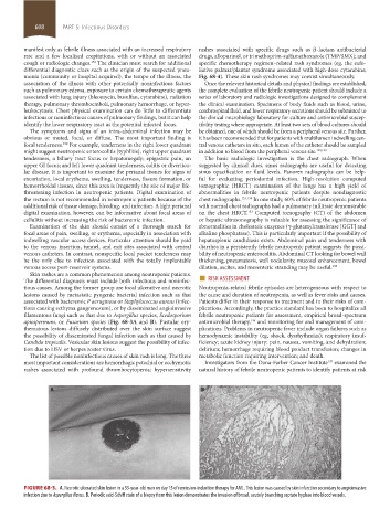
tions causing ecthyma gangrenosum), or by disseminated angioinvasive plications. Accordingly, the practice standard has been to hospitalize all
filamentous fungi such as that due to Aspergillus species, Scedosporium febrile neutropenic patients for assessment, empirical broad-spectrum
apiospermum, or Fusarium species (Fig. 68-3A and B). Pustular ery- antimicrobial therapy, and monitoring for and management of com-
136
thematous lesions diffusely distributed over the skin surface suggest plications. Problems in neutropenic fever include organ failures such as
the possibility of disseminated fungal infection such as that caused by hemodynamic instability (eg, shock, dysrhythmias); respiratory insuf-
Candida tropicalis. Vesicular skin lesions suggest the possibility of infec- ficiency; acute kidney injury; pain, nausea, vomiting, and dehydration;
tion due to HSV or herpes zoster virus. delirium; hemorrhage requiring blood product transfusion; changes in
The list of possible noninfectious causes of skin rash is long. The three metabolic function requiring intervention; and death.
most important considerations are hemorrhagic petechial or ecchymotic Investigators from the Dana-Farber Cancer Institute examined the
137
rashes associated with profound thrombocytopenia; hypersensitivity natural history of febrile neutropenic patients to identify patients at risk
A B
FIGURE 68-3. A. Necrotic ulcerated skin lesion in a 53-year-old man on day 15 of remission-induction therapy for AML. This lesion was caused by skin infarction secondary to angioinvasive
infection due to Aspergillus flavus. B. Periodic acid-Schiff stain of a biopsy from this lesion demonstrates the invasion of broad, acutely branching septate hyphae into blood vessels.
section05_c61-73.indd 608 1/23/2015 12:48:06 PM