
Page 101 - Clinical Anatomy
P. 101
ECA2 7/18/06 6:42 PM Page 86
86 The abdomen and pelvis
component of the external anal sphincter posteriorly into the coccyx;
between the two limbs of the V thus formed, the mucosa is relatively
unsupported and may therefore be torn by a hard faecal mass at this site.
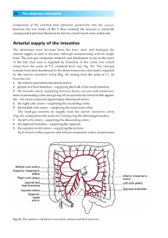
Arterial supply of the intestine
The alimentary tract develops from the fore-, mid- and hind-gut; the
arterial supply to each is discrete, although anastomosing with its neigh-
bour. The fore-gut comprises stomach and duodenum as far as the entry
of the bile duct and is supplied by branches of the coeliac axis which
arises from the aorta at T12 vertebral level (see Fig. 53). The mid-gut
extends from mid-duodenum to the distal transverse colon and is supplied
by the superior mesenteric artery (Fig. 66) arising from the aorta at L1. Its
branches are:
1◊◊the inferior pancreaticoduodenal artery;
2◊◊jejunal and ileal branches—supplying the bulk of the small intestine;
3◊◊the ileocolic artery, supplying terminal ileum, caecum and commence-
ment of ascending colon and giving off an appendicular branch to the appen-
dix—the most commonly ligated intra-abdominal artery;
4◊◊the right colic artery—supplying the ascending colon;
5◊◊the middle colic artery—supplying the transverse colon.
The hind-gut receives its supply from the inferior mesenteric artery
(Fig. 66), arising from the aorta at L3 and giving the following branches:
1◊◊the left colic artery—supplying the descending colon;
2◊◊the sigmoid branches—supplying the sigmoid;
3◊◊the superior rectal artery—supplying the rectum.
Each branch of the superior and inferior mesenteric artery anastomoses
Fig. 66◊The superior and inferior mesenteric arteries and their branches.