
Page 98 - Clinical Anatomy
P. 98
ECA2 7/18/06 6:42 PM Page 83
The gastrointestinal tract 83
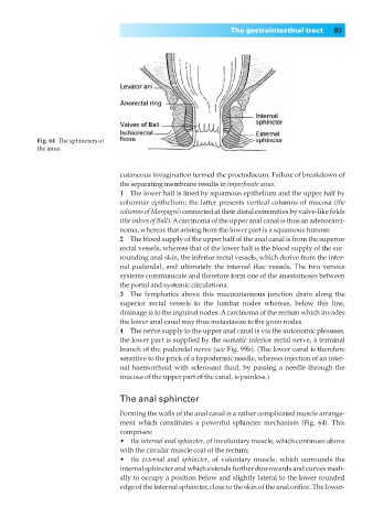
Fig. 64◊The sphincters of
the anus.
cutaneous invagination termed the proctodaeum. Failure of breakdown of
the separating membrane results in imperforate anus.
1◊◊The lower half is lined by squamous epithelium and the upper half by
columnar epithelium; the latter presents vertical columns of mucosa (the
columns of Morgagni) connected at their distal extremities by valve-like folds
(the valves of Ball). Acarcinoma of the upper anal canal is thus an adenocarci-
noma, whereas that arising from the lower part is a squamous tumour.
2◊◊The blood supply of the upper half of the anal canal is from the superior
rectal vessels, whereas that of the lower half is the blood supply of the sur-
rounding anal skin, the inferior rectal vessels, which derive from the inter-
nal pudendal, and ultimately the internal iliac vessels. The two venous
systems communicate and therefore form one of the anastomoses between
the portal and systemic circulations.
3◊◊The lymphatics above this mucocutaneous junction drain along the
superior rectal vessels to the lumbar nodes whereas, below this line,
drainage is to the inguinal nodes. Acarcinoma of the rectum which invades
the lower anal canal may thus metastasize to the groin nodes.
4◊◊The nerve supply to the upper anal canal is via the autonomic plexuses,
the lower part is supplied by the somatic inferior rectal nerve, a terminal
branch of the pudendal nerve (see Fig. 99b). (The lower canal is therefore
sensitive to the prick of a hypodermic needle, whereas injection of an inter-
nal haemorrhoid with sclerosant fluid, by passing a needle through the
mucosa of the upper part of the canal, is painless.)
The anal sphincter
Forming the walls of the anal canal is a rather complicated muscle arrange-
ment which constitutes a powerful sphincter mechanism (Fig. 64). This
comprises:
•◊◊the internal anal sphincter, of involuntary muscle, which continues above
with the circular muscle coat of the rectum;
•◊◊the external anal sphincter, of voluntary muscle, which surrounds the
internal sphincter and which extends further downwards and curves medi-
ally to occupy a position below and slightly lateral to the lower rounded
edge of the internal sphincter, close to the skin of the anal orifice. The lower-