
Page 104 - Clinical Anatomy
P. 104
ECA2 7/18/06 6:42 PM Page 89
The gastrointestinal tract 89
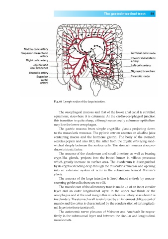
Fig. 68◊Lymph nodes of the large intestine.
The oesophageal mucosa and that of the lower anal canal is stratified
squamous; elsewhere it is columnar. At the cardio-oesophageal junction
this transition is quite sharp, although occasionally columnar epithelium
may line the lower oesophagus.
The gastric mucosa bears simple crypt-like glands projecting down
to the muscularis mucosae. The pyloric antrum secretes an alkaline juice
containing mucus and the hormone gastrin. The body of the stomach
secretes pepsin and also HCl, the latter from the oxyntic cells lying sand-
wiched deeply between the surface cells. The stomach mucosa also pro-
duces intrinsic factor.
The mucosa of the duodenum and small intestine, as well as bearing
crypt-like glands, projects into the bowel lumen in villous processes
which greatly increase its surface area. The duodenum is distinguished
by its crypts extending deep through the muscularis mucosae and opening
into an extensive system of acini in the submucosa termed Brunner’s
glands.
The mucosa of the large intestine is lined almost entirely by mucus-
secreting goblet cells; there are no villi.
The muscle coat of the alimentary tract is made up of an inner circular
layer and an outer longitudinal layer. In the upper two-thirds of the
oesophagus and at the anal margin this muscle is voluntary; elsewhere it is
involuntary. The stomach wall is reinforced by an innermost oblique coat of
muscle and the colon is characterized by the condensation of its longitudi-
nal layer into three taeniae coli.
The autonomic nerve plexuses of Meissner and Auerbach lie respec-
tively in the submucosal layer and between the circular and longitudinal
muscle coats.