
Page 145 - Clinical Anatomy
P. 145
ECA2 7/18/06 6:43 PM Page 130
130 The abdomen and pelvis
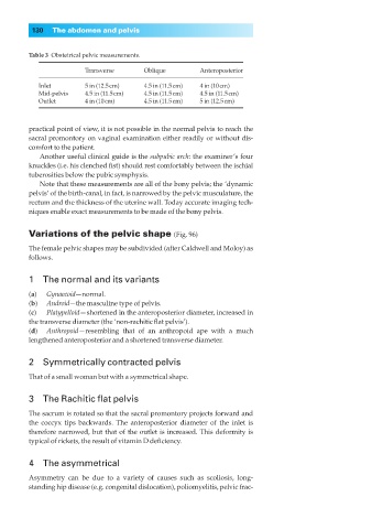
Table 3◊Obstetrical pelvic measurements.
Transverse Oblique Anteroposterior
Inlet 5 in (12.5cm) 4.5 in (11.5cm) 4 in (10cm)
Mid-pelvis 4.5 in (11.5cm) 4.5 in (11.5cm) 4.5 in (11.5cm)
Outlet 4 in (10cm) 4.5 in (11.5cm) 5 in (12.5cm)
practical point of view, it is not possible in the normal pelvis to reach the
sacral promontory on vaginal examination either readily or without dis-
comfort to the patient.
Another useful clinical guide is the subpubic arch: the examiner’s four
knuckles (i.e. his clenched fist) should rest comfortably between the ischial
tuberosities below the pubic symphysis.
Note that these measurements are all of the bony pelvis; the ‘dynamic
pelvis’ of the birth-canal, in fact, is narrowed by the pelvic musculature, the
rectum and the thickness of the uterine wall. Today accurate imaging tech-
niques enable exact measurements to be made of the bony pelvis.
Variations of the pelvic shape (Fig. 96)
The female pelvic shapes may be subdivided (after Caldwell and Moloy) as
follows.
1◊◊The normal and its variants
(a) Gynaecoid—normal.
(b) Android—the masculine type of pelvis.
(c) Platypelloid— shortened in the anteroposterior diameter, increased in
the transverse diameter (the ‘non-rachitic flat pelvis’).
(d) Anthropoid—resembling that of an anthropoid ape with a much
lengthened anteroposterior and a shortened transverse diameter.
2◊◊Symmetrically contracted pelvis
That of a small woman but with a symmetrical shape.
3◊◊The Rachitic flat pelvis
The sacrum is rotated so that the sacral promontory projects forward and
the coccyx tips backwards. The anteroposterior diameter of the inlet is
therefore narrowed, but that of the outlet is increased. This deformity is
typical of rickets, the result of vitamin D deficiency.
4◊◊The asymmetrical
Asymmetry can be due to a variety of causes such as scoliosis, long-
standing hip disease (e.g. congenital dislocation), poliomyelitis, pelvic frac-