
Page 161 - Clinical Anatomy
P. 161
ECA2 7/18/06 6:43 PM Page 146
146 The abdomen and pelvis
Structure
The ovary has no peritoneal covering; the serosa ends at the mesovarian
attachment. It consists of a connective tissue stroma containing Graafian fol-
licles at various stages of development, corpora lutea and corpora albicantia
(hyalinized, regressing corpora lutea, which take several months to absorb
completely).
The surface of the ovary in young children is covered with a so-called
‘germinal epithelium’ of cuboidal cells. It is now known, however, that
the primordial follicles develop in the ovary in early fetal life and do not
differentiate from these cells. In adult life, in fact, the epithelial covering
of the ovary disappears, leaving only a fibrous capsule termed the tunica
albuginea.
After the menopause the ovary becomes small and shrivelled; in old age
the follicles disappear completely.
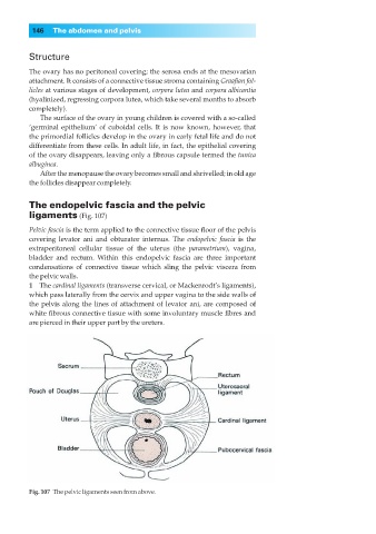
The endopelvic fascia and the pelvic
ligaments (Fig. 107)
Pelvic fascia is the term applied to the connective tissue floor of the pelvis
covering levator ani and obturator internus. The endopelvic fascia is the
extraperitoneal cellular tissue of the uterus (the parametrium), vagina,
bladder and rectum. Within this endopelvic fascia are three important
condensations of connective tissue which sling the pelvic viscera from
the pelvic walls.
1◊◊The cardinal ligaments (transverse cervical, or Mackenrodt’s ligaments),
which pass laterally from the cervix and upper vagina to the side walls of
the pelvis along the lines of attachment of levator ani, are composed of
white fibrous connective tissue with some involuntary muscle fibres and
are pierced in their upper part by the ureters.
Fig. 107◊The pelvic ligaments seen from above.