
Page 164 - Clinical Anatomy
P. 164
ECA2 7/18/06 6:43 PM Page 149
The posterior abdominal wall 149
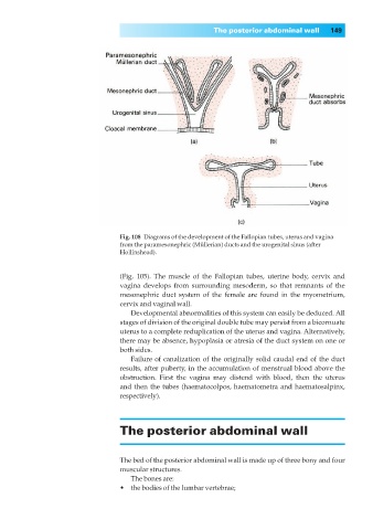
Fig. 108◊Diagrams of the development of the Fallopian tubes, uterus and vagina
from the paramesonephric (Müllerian) ducts and the urogenital sinus (after
Hollinshead).
(Fig. 105). The muscle of the Fallopian tubes, uterine body, cervix and
vagina develops from surrounding mesoderm, so that remnants of the
mesonephric duct system of the female are found in the myometrium,
cervix and vaginal wall.
Developmental abnormalities of this system can easily be deduced. All
stages of division of the original double tube may persist from a bicornuate
uterus to a complete reduplication of the uterus and vagina. Alternatively,
there may be absence, hypoplasia or atresia of the duct system on one or
both sides.
Failure of canalization of the originally solid caudal end of the duct
results, after puberty, in the accumulation of menstrual blood above the
obstruction. First the vagina may distend with blood, then the uterus
and then the tubes (haematocolpos, haematometra and haematosalpinx,
respectively).
The posterior abdominal wall
The bed of the posterior abdominal wall is made up of three bony and four
muscular structures.
The bones are:
•◊◊the bodies of the lumbar vertebrae;