
Page 165 - Clinical Anatomy
P. 165
ECA2 7/18/06 6:43 PM Page 150
150 The abdomen and pelvis
•◊◊the sacrum;
•◊◊the wings of the ilium.
The muscles are:
•◊◊the diaphragm—posterior part;
•◊◊the quadratus lumborum;
•◊◊the psoas major;
•◊◊the iliacus.
The diaphragm has been considered in the section on thorax.
The psoas must be dealt with in more detail because of the involvement
of its sheath in the formation of a psoas abscess.
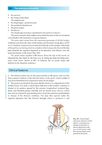
The psoas major arises from the transverse processes of all the lumbar
vertebrae and from the sides of the bodies and the intervening discs of T12
to L5 vertebrae. It passes downwards and laterally at the margin of the brim
of the pelvis, narrowing down to a tendon which crosses the front of the hip
joint beneath the inguinal ligament to be inserted, with iliacus, into the
lesser trochanter of the femur (Fig. 109).
The psoas major, together with iliacus, flexes the hip on the trunk, or,
alternatively, the trunk on the hips (e.g. in sitting up from the lying posi-
tion). Psoas minor, absent in 40% of subjects, lies on psoas major and
attaches to the iliopubic eminence.
Clinical features
1◊◊The femoral artery lies on the psoas tendon in the groin, and it is this
firm posterior relation of the femoral artery at the groin which enables it
here to be identified and compressed easily by the finger.
2◊◊The psoas is enclosed in the psoas sheath which is a compartment of the
lumbar fascia. Pus from a tuberculous infection of the lumbar vertebrae is
limited in its anterior spread by the anterior longitudinal vertebral liga-
ment, and therefore passes laterally into its sheath (psoas abscess), which
may also be entered by pus tracking down from the posterior mediastinum
in disease of the thoracic vertebrae. Pus may then spread under the
inguinal ligament into the femoral triangle where it produces a soft
Fig. 109◊Psoas sheath
and psoas abscess. On the
right is a normal psoas
sheath; on the left it is
shown distended with
pus, which tracks under
the inguinal ligament to
present in the groin.