
Page 241 - Clinical Anatomy
P. 241
ECA4 7/18/06 6:47 PM Page 226
226 The lower limb
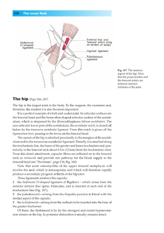
Fig. 167◊The anterior
aspect of the hip. Note
that the psoas tendon and
the femoral artery are
intimate anterior
relations of the joint.
The hip (Figs 166, 167)
The hip is the largest joint in the body. To the surgeon, the examiner and,
therefore, the student it is also the most important.
It is a perfect example of a ball-and-socket joint. Its articular surfaces are
the femoral head and the horse-shoe shaped articular surface of the acetab-
ulum, which is deepened by the fibrocartilaginous labrum acetabulare. The
non-articular lower part of the acetabulum, the acetabular notch, is closed off
below by the transverse acetabular ligament. From this notch is given off the
ligamentum teres, passing to the fovea on the femoral head.
The capsule of the hip is attached proximally to the margins of the acetab-
ulum and to the transverse acetabular ligament. Distally, it is attached along
the trochanteric line, the bases of the greater and lesser trochanters and, pos-
teriorly, to the femoral neck about 0.5in (12mm) from the trochanteric crest.
From this distal attachment, capsular fibres are reflected on to the femoral
neck as retinacula and provide one pathway for the blood supply to the
femoral head (see ‘The femur’, page 216; Fig. 160).
Note that acute osteomyelitis of the upper femoral metaphysis will
involve the neck which is intracapsular and which will therefore rapidly
produce a secondary pyogenic arthritis of the hip joint.
Three ligaments reinforce the capsule:
1◊◊the iliofemoral (Y-shaped ligament of Bigelow) — which arises from the
anterior inferior iliac spine, bifurcates, and is inserted at each end of the
trochanteric line (Fig. 167);
2◊◊the pubofemoral—arising from the iliopubic junction to blend with the
medial aspect of the capsule;
3◊◊the ischiofemoral—arising from the ischium to be inserted into the base of
the greater trochanter.
Of these, the iliofemoral is by far the strongest and resists hyperexten-
sion strains on the hip. In posterior dislocation it usually remains intact.