
Page 237 - Clinical Anatomy
P. 237
ECA4 7/18/06 6:47 PM Page 222
222 The lower limb
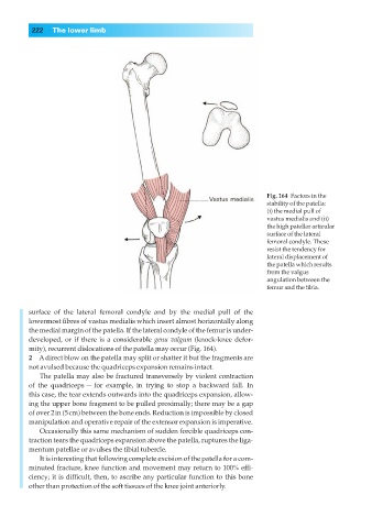
Fig. 164◊Factors in the
stability of the patella:
(i) the medial pull of
vastus medialis and (ii)
the high patellar articular
surface of the lateral
femoral condyle. These
resist the tendency for
lateral displacement of
the patella which results
from the valgus
angulation between the
femur and the tibia.
surface of the lateral femoral condyle and by the medial pull of the
lowermost fibres of vastus medialis which insert almost horizontally along
the medial margin of the patella. If the lateral condyle of the femur is under-
developed, or if there is a considerable genu valgum (knock-knee defor-
mity), recurrent dislocations of the patella may occur (Fig. 164).
2◊◊A direct blow on the patella may split or shatter it but the fragments are
not avulsed because the quadriceps expansion remains intact.
The patella may also be fractured transversely by violent contraction
of the quadriceps — for example, in trying to stop a backward fall. In
this case, the tear extends outwards into the quadriceps expansion, allow-
ing the upper bone fragment to be pulled proximally; there may be a gap
of over 2in (5cm) between the bone ends. Reduction is impossible by closed
manipulation and operative repair of the extensor expansion is imperative.
Occasionally this same mechanism of sudden forcible quadriceps con-
traction tears the quadriceps expansion above the patella, ruptures the liga-
mentum patellae or avulses the tibial tubercle.
It is interesting that following complete excision of the patella for a com-
minuted fracture, knee function and movement may return to 100% effi-
ciency; it is difficult, then, to ascribe any particular function to this bone
other than protection of the soft tissues of the knee joint anteriorly.