
Page 25 - Clinical Anatomy
P. 25
ECA1 7/18/06 6:31 PM Page 10
10 The Thorax
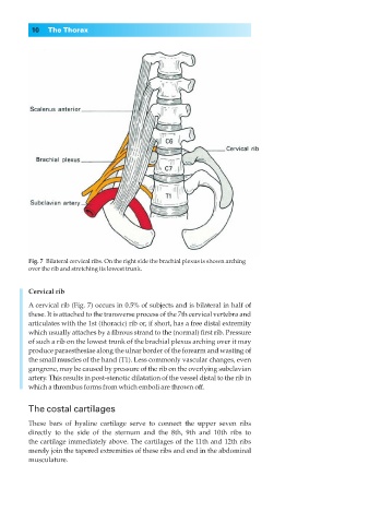
Fig. 7◊Bilateral cervical ribs. On the right side the brachial plexus is shown arching
over the rib and stretching its lowest trunk.
Cervical rib
A cervical rib (Fig. 7) occurs in 0.5% of subjects and is bilateral in half of
these. It is attached to the transverse process of the 7th cervical vertebra and
articulates with the 1st (thoracic) rib or, if short, has a free distal extremity
which usually attaches by a fibrous strand to the (normal) first rib. Pressure
of such a rib on the lowest trunk of the brachial plexus arching over it may
produce paraesthesiae along the ulnar border of the forearm and wasting of
the small muscles of the hand (T1). Less commonly vascular changes, even
gangrene, may be caused by pressure of the rib on the overlying subclavian
artery. This results in post-stenotic dilatation of the vessel distal to the rib in
which a thrombus forms from which emboli are thrown off.
The costal cartilages
These bars of hyaline cartilage serve to connect the upper seven ribs
directly to the side of the sternum and the 8th, 9th and 10th ribs to
the cartilage immediately above. The cartilages of the 11th and 12th ribs
merely join the tapered extremities of these ribs and end in the abdominal
musculature.