
Page 27 - Clinical Anatomy
P. 27
ECA1 7/18/06 6:31 PM Page 12
12 The Thorax
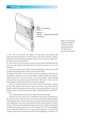
Fig. 8◊The relationship
of an intercostal space.
(Note that a needle
passed into the chest
immediately above a
rib will avoid the
neurovascular bundle.)
1◊◊the external intercostal, the fibres of which pass downwards and
forwards from the rib above to the rib below and reach from the vertebrae
behind to the costochondral junction in front, where muscle is replaced by
the anterior intercostal membrane;
2◊◊the internal intercostal, which runs downwards and backwards from the
sternum to the angles of the ribs where it becomes the posterior intercostal
membrane;
3◊◊the innermost intercostal, which is only incompletely separated from the
internal intercostal muscle by the neurovascular bundle.
The fibres of this sheet cross more than one intercostal space and it may be
incomplete. Anteriorly it has a more distinct portion which is fan-like in
shape, termed the transversus thoracis (or sternocostalis), which spreads
upwards from the posterior aspect of the lower sternum to insert onto the
inner surfaces of the second to the sixth costal cartilages.
Just as in the abdomen, the nerves and vessels of the thoracic wall lie
between the middle and innermost layers of muscles. This neurovascular
bundle consists, from above downwards, of vein, artery and nerve, the vein
lying in a groove on the undersurface of the corresponding rib (remember—
v,a,n).
The vessels comprise the posterior and anterior intercostals.
The posterior intercostal arteries of the lower nine spaces are branches of
the thoracic aorta, while the first two are derived from the superior inter-
costal branch of the costocervical trunk, the only branch of the second part
of the subclavian artery. Each runs forward in the subcostal groove to anas-
tomose with the anterior intercostal artery. Each has a number of branches
to adjacent muscles, to the skin and to the spinal cord. The corresponding
veins are mostly tributaries of the azygos and hemiazygos veins. The first
posterior intercostal vein drains into the brachiocephalic or vertebral vein.