
Page 30 - Clinical Anatomy
P. 30
ECA1 7/18/06 6:31 PM Page 15
The thoracic cage 15
Oesophagus
Inferior vena cava
Left phrenic nerve
Right phrenic nerve Vagi
Right splanchnic Aorta
nerve Left splanchnic
nerve
Subcostal nerve
Transverse abdominis
muscle
Quadratus lumborum
muscle Sympathetic trunk
Psoas major
muscle
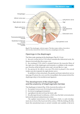
Fig. 10◊The diaphragm—inferior aspect. The three major orifices, from above
downwards, transmit the inferior vena cava, oesophagus and aorta.
Openings in the diaphragm
The three main openings in the diaphragm (Figs 10, 11) are:
1◊◊the aortic (at the level of T12) which transmits the abdominal aorta, the
thoracic duct and often the azygos vein;
2◊◊the oesophageal (T10) which is situated between the muscular fibres of
the right crus of the diaphragm and transmits, in addition to the oesopha-
gus, branches of the left gastric artery and vein and the two vagi;
3◊◊the opening for the inferior vena cava (T8) which is placed in the central
tendon and also transmits the right phrenic nerve.
In addition to these structures, the greater and lesser splanchnic nerves
(see page 49) pierce the crura and the sympathetic chain passes behind the
diaphragm deep to the medial arcuate ligament.
The development of the diaphragm
and the anatomy of diaphragmatic herniae
The diaphragm is formed (Fig. 12) by fusion in the embryo of:
1◊◊the septum transversum (forming the central tendon);
2◊◊the dorsal oesophageal mesentery;
3◊◊a peripheral rim derived from the body wall;
4◊◊the pleuroperitoneal membranes, which close the fetal communication
between the pleural and peritoneal cavities.
The septum transversum is the mesoderm which, in early develop-
ment, lies in front of the head end of the embryo. With the folding off of the
head, this mesodermal mass is carried ventrally and caudally, to lie in its