
Page 28 - Clinical Anatomy
P. 28
ECA1 7/18/06 6:31 PM Page 13
The thoracic cage 13
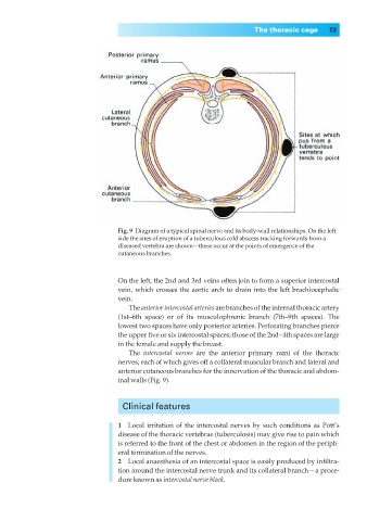
Fig. 9◊Diagram of a typical spinal nerve and its body-wall relationships. On the left
side the sites of eruption of a tuberculous cold abscess tracking forwards from a
diseased vertebra are shown—these occur at the points of emergence of the
cutaneous branches.
On the left, the 2nd and 3rd veins often join to form a superior intercostal
vein, which crosses the aortic arch to drain into the left brachiocephalic
vein.
The anterior intercostal arteries are branches of the internal thoracic artery
(1st–6th space) or of its musculophrenic branch (7th–9th spaces). The
lowest two spaces have only posterior arteries. Perforating branches pierce
the upper five or six intercostal spaces; those of the 2nd–4th spaces are large
in the female and supply the breast.
The intercostal nerves are the anterior primary rami of the thoracic
nerves, each of which gives off a collateral muscular branch and lateral and
anterior cutaneous branches for the innervation of the thoracic and abdom-
inal walls (Fig. 9).
Clinical features
1◊◊Local irritation of the intercostal nerves by such conditions as Pott’s
disease of the thoracic vertebrae (tuberculosis) may give rise to pain which
is referred to the front of the chest or abdomen in the region of the periph-
eral termination of the nerves.
2◊◊Local anaesthesia of an intercostal space is easily produced by infiltra-
tion around the intercostal nerve trunk and its collateral branch— a proce-
dure known as intercostal nerve block.