
Page 262 - Clinical Anatomy
P. 262
ECA4 7/18/06 6:47 PM Page 247
The veins of the lower limb 247
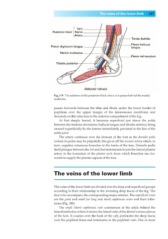
Fig. 179◊The relations of the posterior tibial artery as it passes behind the medial
malleolus.
passes forwards between the tibia and fibula under the lower border of
popliteus over the upper margin of the interosseous membrane and
descends on this structure in the anterior compartment of the leg.
At first deeply buried, it becomes superficial just above the ankle
between the tendons of extensor hallucis longus and tibialis anterior, being
crossed superficially by the former immediately proximal to the line of the
ankle joint.
The artery continues over the dorsum of the foot as the dorsalis pedis
(where its pulse may be palpated); this gives off the arcuate artery which, in
turn, supplies cutaneous branches to the backs of the toes. Dorsalis pedis
itself plunges between the 1st and 2nd metatarsals to join the lateral plantar
artery in the formation of the plantar arch, from which branches run for-
wards to supply the plantar aspects of the toes.
The veins of the lower limb
The veins of the lower limb are divided into the deep and superficial groups
according to their relationship to the investing deep fascia of the leg. The
deep veins accompany the corresponding major arteries. The superficial veins
are the great and small (or long and short) saphenous veins and their tribu-
taries (Fig. 180).
The small (short) saphenous vein commences at the ankle behind the
lateral malleolus where it drains the lateral side of the dorsal venous plexus
of the foot. It courses over the back of the calf, perforates the deep fascia
over the popliteal fossa and terminates in the popliteal vein. One or more