
Page 265 - Clinical Anatomy
P. 265
ECA4 7/18/06 6:47 PM Page 250
250 The lower limb
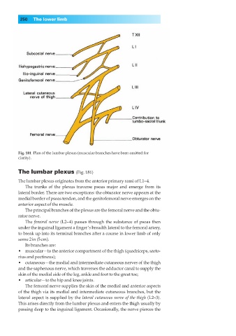
Fig. 181◊Plan of the lumbar plexus (muscular branches have been omitted for
clarity).
The lumbar plexus (Fig. 181)
The lumbar plexus originates from the anterior primary rami of L1–4.
The trunks of the plexus traverse psoas major and emerge from its
lateral border. There are two exceptions: the obturator nerve appears at the
medial border of psoas tendon, and the genitofemoral nerve emerges on the
anterior aspect of the muscle.
The principal branches of the plexus are the femoral nerve and the obtu-
rator nerve.
The femoral nerve (L2–4) passes through the substance of psoas then
under the inguinal ligament a finger’s breadth lateral to the femoral artery,
to break up into its terminal branches after a course in lower limb of only
some 2in (5cm).
Its branches are:
•◊◊muscular—to the anterior compartment of the thigh (quadriceps, sarto-
rius and pectineus);
•◊◊cutaneous—the medial and intermediate cutaneous nerves of the thigh
and the saphenous nerve, which traverses the adductor canal to supply the
skin of the medial side of the leg, ankle and foot to the great toe;
•◊◊articular—to the hip and knee joints.
The femoral nerve supplies the skin of the medial and anterior aspects
of the thigh via its medial and intermediate cutaneous branches, but the
lateral aspect is supplied by the lateral cutaneous nerve of the thigh (L2–3).
This arises directly from the lumbar plexus and enters the thigh usually by
passing deep to the inguinal ligament. Occasionally, the nerve pierces the