
Page 267 - Clinical Anatomy
P. 267
ECA4 7/18/06 6:47 PM Page 252
252 The lower limb
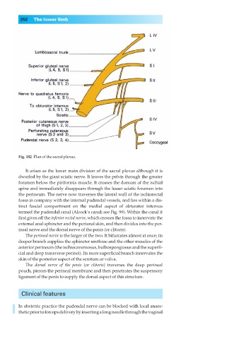
Fig. 182◊Plan of the sacral plexus.
It arises as the lower main division of the sacral plexus although it is
dwarfed by the giant sciatic nerve. It leaves the pelvis through the greater
foramen below the piriformis muscle. It crosses the dorsum of the ischial
spine and immediately disappears through the lesser sciatic foramen into
the perineum. The nerve now traverses the lateral wall of the ischiorectal
fossa in company with the internal pudendal vessels, and lies within a dis-
tinct fascial compartment on the medial aspect of obturator internus
termed the pudendal canal (Alcock’s canal; see Fig. 99). Within the canal it
first gives off the inferior rectal nerve, which crosses the fossa to innervate the
external anal sphincter and the perianal skin, and then divides into the per-
ineal nerve and the dorsal nerve of the penis (or clitoris).
The perineal nerve is the larger of the two. It bifurcates almost at once; its
deeper branch supplies the sphincter urethrae and the other muscles of the
anterior perineum (the ischiocavernosus, bulbospongiosus and the superfi-
cial and deep transverse perinei). Its more superficial branch innervates the
skin of the posterior aspect of the scrotum or vulva.
The dorsal nerve of the penis (or clitoris) traverses the deep perineal
pouch, pierces the perineal membrane and then penetrates the suspensory
ligament of the penis to supply the dorsal aspect of this structure.
Clinical features
In obstetric practice the pudendal nerve can be blocked with local anaes-
thetic prior to forceps delivery by inserting a long needle through the vaginal