
Page 257 - Clinical Anatomy
P. 257
ECA4 7/18/06 6:47 PM Page 242
242 The lower limb
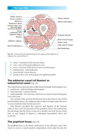
Fig. 177◊Cross-section through the thigh in the region of the adductor, or
subsartorial, canal of Hunter.
•◊◊artery—aneurysm of the femoral artery;
•◊◊vein—varix of the great saphenous vein;
•◊◊nerve—neuroma of the femoral nerve or its branches;
•◊◊femoral canal—femoral hernia;
•◊◊psoas sheath—psoas abscess;
•◊◊lymph nodes—any of the causes of lymphadenopathy.
The adductor canal (of Hunter) or
subsartorial canal (Fig. 177)
This canal leads on from the apex of the femoral triangle. Its boundaries are:
•◊◊posteriorly—adductor longus and magnus;
•◊◊anterolaterally—vastus medialis;
•◊◊anteromedially—the sartorius, which lies on a fascial sheet forming the
roof of the canal.
The contents of the canal are the femoral artery, the femoral vein (which
lies behind the artery), the saphenous nerve and, in its upper part, the nerve
to vastus medialis from the femoral nerve.
John Hunter described the exposure and ligation of the femoral
artery in this canal for aneurysm of the popliteal artery; this method has
the advantage that the artery at this site is healthy and will not tear when
tied, as may happen if ligation is attempted immediately above the
aneurysm.
The popliteal fossa (Fig. 178)
The popliteal fossa is the distal continuation of the adductor canal. This
‘fossa’ is, in fact, a closely packed compartment which only becomes the