
Page 278 - Clinical Anatomy
P. 278
ECA5 7/18/06 6:50 PM Page 263
The surface anatomy of the neck 263
Anterior jugular
Pretracheal fascia
(containing thyroid, vein
trachea, oesophagus
and recurrent nerve)
Investing fascia
Sternocleidomastoid
Sternohyoid
Sternothyroid
Omohyoid
External jugular vein
C6 Carotid sheath (containing
Pre-vertebral fascia common carotid artery,
internal jugular vein, and
vagus nerve) with sympathetic
chain behind (a)
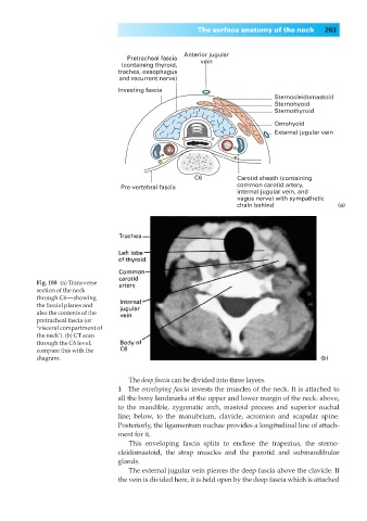
Fig. 188◊(a) Transverse
section of the neck
through C6—showing
the fascial planes and
also the contents of the
pretracheal fascia (or
‘visceral compartment of
the neck’). (b) CT scan
through the C6 level;
compare this with the
diagram. (b)
The deep fascia can be divided into three layers.
1◊◊The enveloping fascia invests the muscles of the neck. It is attached to
all the bony landmarks at the upper and lower margin of the neck: above,
to the mandible, zygomatic arch, mastoid process and superior nuchal
line; below, to the manubrium, clavicle, acromion and scapular spine.
Posteriorly, the ligamentum nuchae provides a longitudinal line of attach-
ment for it.
This enveloping fascia splits to enclose the trapezius, the sterno-
cleidomastoid, the strap muscles and the parotid and submandibular
glands.
The external jugular vein pierces the deep fascia above the clavicle. If
the vein is divided here, it is held open by the deep fascia which is attached