
Page 283 - Clinical Anatomy
P. 283
ECA5 7/18/06 6:50 PM Page 268
268 The head and neck
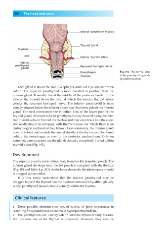
Fig. 192◊The normal sites
of the parathyroid glands
(posterior aspect).
Each gland is about the size of a split pea and is of a yellowish-brown
colour. The superior parathyroid is more constant in position than the
inferior gland. It usually lies at the middle of the posterior border of the
lobe of the thyroid above the level at which the inferior thyroid artery
crosses the recurrent laryngeal nerve. The inferior parathyroid is most
usually situated below the inferior artery near the lower pole of the thyroid
gland. The next commonest site is within 1cm of the lower pole of the
thyroid gland. Aberrant inferior parathyroids may descend along the infe-
rior thyroid veins in front of the trachea and may even track into the supe-
rior mediastinum in company with thymic tissues, for which there is an
embryological explanation (see below). Less commonly, the inferior gland
may lie behind and outside the fascial sheath of the thyroid and be found
behind the oesophagus or even in the posterior mediastinum. Only on
extremely rare occasions are the glands actually completely buried within
thyroid tissue (Fig. 193).
Development
The superior parathyroids differentiate from the 4th branchial pouch. The
inferior gland develops from the 3rd pouch in company with the thymus
(Fig. 194 and Table 4, p. 311). As the latter descends, the inferior parathyroid
is dragged down with it.
It is thus easily understood that the inferior parathyroid may be
dragged beyond the thyroid into the mediastinum and why, although very
rarely, parathyroid tissue is found actually within the thymus.
Clinical features
1◊◊These possible aberrant sites are, of course, of great importance in
searching for a parathyroid adenoma in hyperparathyroidism.
2◊◊The parathyroids are usually safe in subtotal thyroidectomy because
the posterior rim of the thyroid is preserved. However, they may be