
Page 282 - Clinical Anatomy
P. 282
ECA5 7/18/06 6:50 PM Page 267
The thyroid gland 267
chain may be involved, producing changes in the voice and Horner’s syn-
drome respectively.
3◊◊We have already noted, in dealing with the fasciae of the neck, that the
thyroid gland is enclosed in the pretracheal fascia. This thyroid capsule is
much denser in front than behind and the enlarging gland therefore tends
to push backwards, burying itself round the sides and even the back of the
trachea and oesophagus. Because of the attachments of its fascial com-
partment, a large goitre will also extend downwards into the superior
mediastinum (‘plunging goitre’).
Above, the pretracheal fascia blends with the larynx, accounting for the
upward movement of the thyroid gland with each act of swallowing.
4◊◊Thyroidectomy is carried out through a transverse ‘collar’ incision, two
fingers’ breadth above the suprasternal notch. This lies in the line of the
natural skin folds of the neck. Skin flaps are reflected, together with
platysma, and the investing fascia opened longitudinally between the strap
muscles and between the anterior jugular veins.
If more room is required in the case of a large goitre, the strap muscles
are divided; this is carried out at their upper extremity because their nerve
supply (the ansa hypoglossi) enters the lower part of the muscles and is
hence preserved.
The pretracheal fascia is then divided, exposing the thyroid gland;
unless this tissue plane deep to the fascia is found, dissection is a difficult
and bloody procedure.
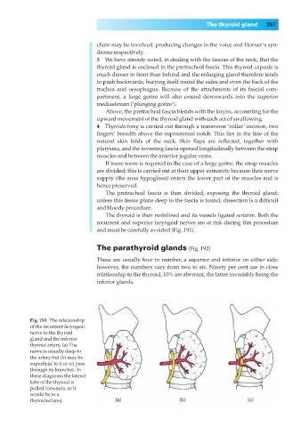
The thyroid is then mobilized and its vessels ligated seriatim. Both the
recurrent and superior laryngeal nerves are at risk during this procedure
and must be carefully avoided (Fig. 191).
The parathyroid glands (Fig. 192)
These are usually four in number, a superior and inferior on either side;
however, the numbers vary from two to six. Ninety per cent are in close
relationship to the thyroid, 10% are aberrant, the latter invariably being the
inferior glands.
Fig. 191◊The relationship
of the recurrent laryngeal
nerve to the thyroid
gland and the inferior
thyroid artery. (a) The
nerve is usually deep to
the artery but (b) may be
superficial to it or (c) pass
through its branches. In
these diagrams the lateral
lobe of the thyroid is
pulled forwards, as it
would be in a
thyroidectomy.