
Page 390 - Clinical Anatomy
P. 390
ECA6 7/18/06 6:54 PM Page 375
The cranial nerves 375
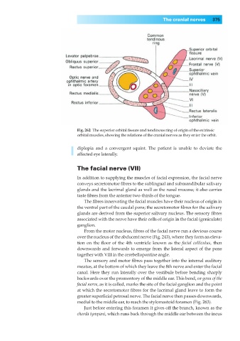
Fig. 262◊The superior orbital fissure and tendinous ring of origin of the extrinsic
orbital muscles, showing the relations of the cranial nerves as they enter the orbit.
diplopia and a convergent squint. The patient is unable to deviate the
affected eye laterally.
The facial nerve (VII)
In addition to supplying the muscles of facial expression, the facial nerve
conveys secretomotor fibres to the sublingual and submandibular salivary
glands and the lacrimal gland as well as the nasal mucosa; it also carries
taste fibres from the anterior two-thirds of the tongue.
The fibres innervating the facial muscles have their nucleus of origin in
the ventral part of the caudal pons; the secretomotor fibres for the salivary
glands are derived from the superior salivary nucleus. The sensory fibres
associated with the nerve have their cells of origin in the facial (geniculate)
ganglion.
From the motor nucleus, fibres of the facial nerve run a devious course
over the nucleus of the abducent nerve (Fig. 243), where they form an eleva-
tion on the floor of the 4th ventricle known as the facial colliculus, then
downwards and forwards to emerge from the lateral aspect of the pons
together with VIII in the cerebellopontine angle.
The sensory and motor fibres pass together into the internal auditory
meatus, at the bottom of which they leave the 8th nerve and enter the facial
canal. Here they run laterally over the vestibule before bending sharply
backwards over the promontory of the middle ear. This bend, or genu of the
facial nerve, as it is called, marks the site of the facial ganglion and the point
at which the secretomotor fibres for the lacrimal gland leave to form the
greater superficial petrosal nerve. The facial nerve then passes downwards,
medial to the middle ear, to reach the stylomastoid foramen (Fig. 263).
Just before entering this foramen it gives off the branch, known as the
chorda tympani, which runs back through the middle ear between the incus