
Page 385 - Clinical Anatomy
P. 385
ECA6 7/18/06 6:54 PM Page 370
370 The central nervous system
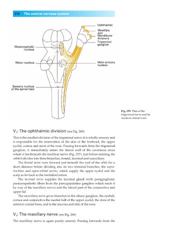
Fig. 259◊Plan of the
trigeminal nerve and its
nuclei in dorsal view.
V : The ophthalmic division (see Fig. 260)
1
This is the smallest division of the trigeminal nerve; it is wholly sensory and
is responsible for the innervation of the skin of the forehead, the upper
eyelid, cornea and most of the nose. Passing forwards from the trigeminal
ganglion, it immediately enters the lateral wall of the cavernous sinus
where it lies beneath the trochlear nerve (Fig. 257). Just before entering the
orbit it divides into three branches, frontal, lacrimal and nasociliary.
The frontal nerve runs forward just beneath the roof of the orbit for a
short distance before dividing into its two terminal branches, the supra-
trochlear and supra-orbital nerves, which supply the upper eyelid and the
scalp as far back as the lambdoid suture.
The lacrimal nerve supplies the lacrimal gland (with postganglionic
parasympathetic fibres from the pterygopalatine ganglion which reach it
by way of the maxillary nerve) and the lateral part of the conjunctiva and
upper lid.
The nasociliary nerve gives branches to the ciliary ganglion, the eyeball,
cornea and conjunctiva the medial half of the upper eyelid, the dura of the
anterior cranial fossa, and to the mucosa and skin of the nose.
V : The maxillary nerve (see Fig. 260)
2
The maxillary nerve is again purely sensory. Passing forwards from the