
Page 389 - Clinical Anatomy
P. 389
ECA6 7/18/06 6:54 PM Page 374
374 The central nervous system
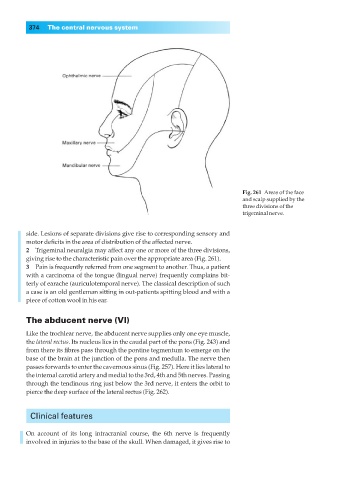
Fig. 261◊Areas of the face
and scalp supplied by the
three divisions of the
trigeminal nerve.
side. Lesions of separate divisions give rise to corresponding sensory and
motor deficits in the area of distribution of the affected nerve.
2◊◊Trigeminal neuralgia may affect any one or more of the three divisions,
giving rise to the characteristic pain over the appropriate area (Fig. 261).
3◊◊Pain is frequently referred from one segment to another. Thus, a patient
with a carcinoma of the tongue (lingual nerve) frequently complains bit-
terly of earache (auriculotemporal nerve). The classical description of such
a case is an old gentleman sitting in out-patients spitting blood and with a
piece of cotton wool in his ear.
The abducent nerve (VI)
Like the trochlear nerve, the abducent nerve supplies only one eye muscle,
the lateral rectus. Its nucleus lies in the caudal part of the pons (Fig. 243) and
from there its fibres pass through the pontine tegmentum to emerge on the
base of the brain at the junction of the pons and medulla. The nerve then
passes forwards to enter the cavernous sinus (Fig. 257). Here it lies lateral to
the internal carotid artery and medial to the 3rd, 4th and 5th nerves. Passing
through the tendinous ring just below the 3rd nerve, it enters the orbit to
pierce the deep surface of the lateral rectus (Fig. 262).
Clinical features
On account of its long intracranial course, the 6th nerve is frequently
involved in injuries to the base of the skull. When damaged, it gives rise to