
Page 397 - Clinical Anatomy
P. 397
ECA6 7/18/06 6:54 PM Page 382
382 The central nervous system
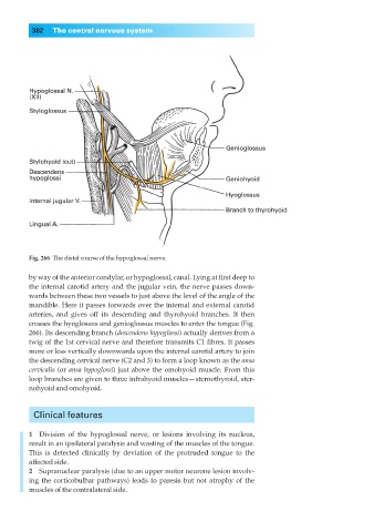
Fig. 266◊The distal course of the hypoglossal nerve.
by way of the anterior condylar, or hypoglossal, canal. Lying at first deep to
the internal carotid artery and the jugular vein, the nerve passes down-
wards between these two vessels to just above the level of the angle of the
mandible. Here it passes forwards over the internal and external carotid
arteries, and gives off its descending and thyrohyoid branches. It then
crosses the hyoglossus and genioglossus muscles to enter the tongue (Fig.
266). Its descending branch (descendens hypoglossi) actually derives from a
twig of the 1st cervical nerve and therefore transmits C1 fibres. It passes
more or less vertically downwards upon the internal carotid artery to join
the descending cervical nerve (C2 and 3) to form a loop known as the ansa
cervicalis (or ansa hypoglossi) just above the omohyoid muscle. From this
loop branches are given to three infrahyoid muscles— sternothyroid, ster-
nohyoid and omohyoid.
Clinical features
1◊◊Division of the hypoglossal nerve, or lesions involving its nucleus,
result in an ipsilateral paralysis and wasting of the muscles of the tongue.
This is detected clinically by deviation of the protruded tongue to the
affected side.
2◊◊Supranuclear paralysis (due to an upper motor neurone lesion involv-
ing the corticobulbar pathways) leads to paresis but not atrophy of the
muscles of the contralateral side.