
Page 402 - Clinical Anatomy
P. 402
ECA6 7/18/06 6:54 PM Page 387
The special senses 387
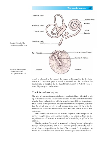
Fig. 269◊Detail of the
membranous labyrinth.
Fig. 270◊The tympanic
membrane as seen
through an auroscope.
which is attached to the neck of the stapes and is supplied by the facial
nerve, and the tensor tympani, which is inserted into the handle of the
malleus and is supplied by the mandibular division of V. Both serve to
damp high-frequency vibrations.
The internal ear (Fig. 269)
The internal ear consists essentially of a complicated bony labyrinth made
up of a central vestibule, which communicates posteriorly with three semi-
circular ducts and anteriorly with the spiral cochlea. This cavity contains a
fluid known as perilymph and encloses the membranous labyrinth, compris-
ing the utricle and saccule, which communicate respectively with the
semicircular canals and the cochlear canal. The duct system is filled with
endolymph.
In each component of the membranous labyrinth there are specialized
sensory receptor areas known as the maculae of the utricle and saccule, the
ampullary crests of the semicircular canals and the spiral organ of Corti in the
cochlea.
The disposition of the semicircular canals in three planes at right angles
to each other renders this part of the labyrinth particularly well suited to
signal changes in position of the head. The organ of Corti is adapted to
record the sound vibrations transmitted by the stapes at the oval window.