
Page 400 - Clinical Anatomy
P. 400
ECA6 7/18/06 6:54 PM Page 385
The special senses 385
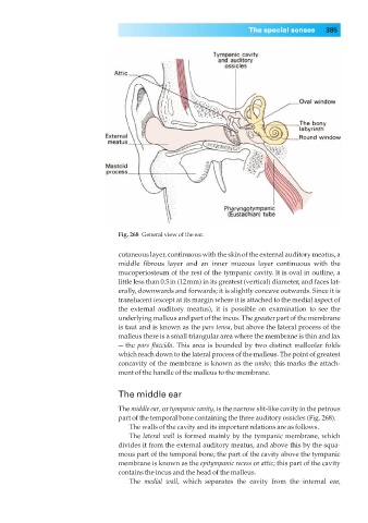
Fig. 268◊General view of the ear.
cutaneous layer, continuous with the skin of the external auditory meatus, a
middle fibrous layer and an inner mucous layer continuous with the
mucoperiosteum of the rest of the tympanic cavity. It is oval in outline, a
little less than 0.5in (12mm) in its greatest (vertical) diameter, and faces lat-
erally, downwards and forwards; it is slightly concave outwards. Since it is
translucent (except at its margin where it is attached to the medial aspect of
the external auditory meatus), it is possible on examination to see the
underlying malleus and part of the incus. The greater part of the membrane
is taut and is known as the pars tensa, but above the lateral process of the
malleus there is a small triangular area where the membrane is thin and lax
— the pars flaccida. This area is bounded by two distinct malleolar folds
which reach down to the lateral process of the malleus. The point of greatest
concavity of the membrane is known as the umbo; this marks the attach-
ment of the handle of the malleus to the membrane.
The middle ear
The middle ear, or tympanic cavity, is the narrow slit-like cavity in the petrous
part of the temporal bone containing the three auditory ossicles (Fig. 268).
The walls of the cavity and its important relations are as follows.
The lateral wall is formed mainly by the tympanic membrane, which
divides it from the external auditory meatus, and above this by the squa-
mous part of the temporal bone; the part of the cavity above the tympanic
membrane is known as the epitympanic recess or attic; this part of the cavity
contains the incus and the head of the malleus.
The medial wall, which separates the cavity from the internal ear,