
Page 415 - Clinical Anatomy
P. 415
ECA6 7/18/06 6:54 PM Page 400
400 The central nervous system
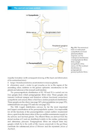
Fig. 278◊The anatomical
basis of widespread
sympathetic and local
parasympathetic
response. (a) The
widespread distribution
of postganglionic fibres
from a single
sympathetic white
ramus. (b) The localized
distribution of
postganglionic
parasympathetic fibres.
impulse formation (with consequent slowing of the heart and diminution
of its contraction force);
5◊◊lungs—bronchoconstrictor, secretomotor to mucous glands;
6◊◊alimentary canal — motor to gut muscles as far as the region of the
ascending colon; inhibitor to the pyloric sphincter; secretomotor to the
glands and adnexae of the stomach and intestine.
The parasympathetic distribution of III, VII and IX is carried out via
four ganglia from which postganglionic fibres relay. These ganglia also
transmit (without synapse and therefore without functional connection)
sympathetic and sensory fibres which have similar peripheral distribution.
These ganglia are the ciliary (see page 367), pterygopalatine (see page 371),
submandibular (see page 373) and otic (see page 372).
The 10th (vagal) distribution conveys by far the most important
and largest contributions of the parasympathetic system. It is responsible
for all the functions of the parasympathetic cranial outflow enumerated
above, apart from the innervation of the eye and the secretomotor supply to
the salivary and lacrimal glands. The efferent fibres are derived from the
dorsal nucleus of X and are distributed widely in the cardiac, pulmonary
and alimentary plexuses. Postganglionic fibres are relayed from tiny
ganglia which lie in the walls of the viscera concerned; in the gut these
constitute the submucosal plexus of Meissner and the myenteric plexus of
Auerbach.