
Page 414 - Clinical Anatomy
P. 414
ECA6 7/18/06 6:54 PM Page 399
The autonomic nervous system 399
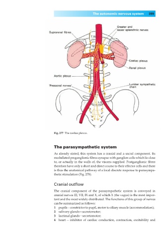
Fig. 277◊The coeliac plexus.
The parasympathetic system
As already stated, this system has a cranial and a sacral component. Its
medullated preganglionic fibres synapse with ganglion cells which lie close
to, or actually in the walls of, the viscera supplied. Postganglionic fibres
therefore have only a short and direct course to their effector cells and there
is thus the anatomical pathway of a local discrete response to parasympa-
thetic stimulation (Fig. 278).
Cranial outflow
The cranial component of the parasympathetic system is conveyed in
cranial nerves III, VII, IX and X, of which X (the vagus) is the most impor-
tant and the most widely distributed. The functions of this group of nerves
can be summarized as follows:
1◊◊pupils—constrictor to pupil, motor to ciliary muscle (accommodation);
2◊◊salivary glands—secretomotor;
3◊◊lacrimal glands—secretomotor;
4◊◊heart — inhibitor of cardiac conduction, contraction, excitability and